

- ¥3500 - 4500
- 欣润生物(NEWGAINBIO)
- 江苏无锡
- IH1001
- 2025年12月15日

企业认证
相关产品推荐更多 >


万千商家帮你免费找货
0 人在求购买到急需产品
- 详细信息
- 询价记录
- 文献和实验
- 技术资料
- 英文名:
Immortalized human skin fibroblasts
- 库存:
100万
- 供应商:
欣润生物
- 肿瘤类型:
否
- 细胞类型:
原代细胞永生化
- ATCC Number:
无
- 品系:
人源
- 组织来源:
皮肤
- 相关疾病:
无
- 物种来源:
人源
- 免疫类型:
不详
- 细胞形态:
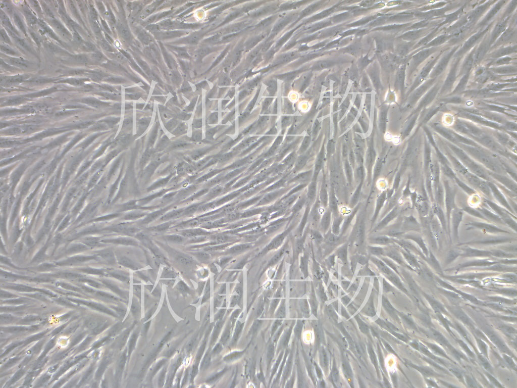
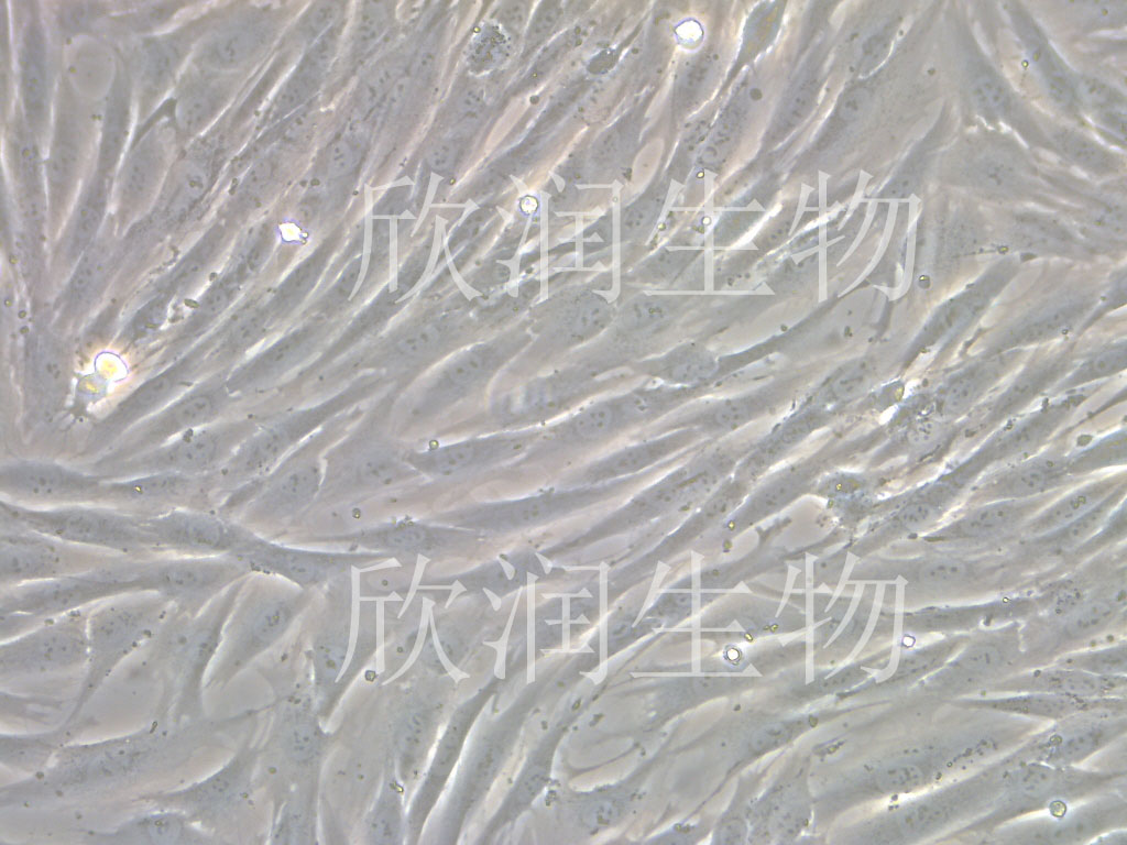
成纤维细胞样贴壁生物
- 是否是肿瘤细胞:
否
- 器官来源:
皮肤
- 运输方式:
常温
- 年限:
青年
- 生长状态:
贴壁生长
- 规格:
T25方瓶
产品介绍
细胞名称:人皮肤成纤维细胞系、永生化人皮肤成纤维细胞、人真皮成纤维细胞
背景描述:人真皮组织主要由成纤维细胞及其产生的纤维、基质构成,并有血管、淋巴管、神经、皮肤附属器及其他细胞成分。人皮肤成纤维细胞系、永生化人皮肤成纤维细胞、人真皮成纤维细胞主要分布于黏膜和皮下疏松结缔组织,胞质内富含嗜碱性颗粒,细胞表面能够表达高亲和力FcεRI,可结合游离IgE,参与I型超敏反应。。
产品货号:IH1001
细胞类型:永生化的细胞
传代能力:传代30代左右
细胞形态:成纤维细胞样
培养基:人皮肤成纤维细胞系、永生化人皮肤成纤维细胞、人真皮成纤维细胞完全培养基
支原体:阴性
培养条件:37℃,5%CO2
发货方式:T25方瓶
货期:1周左右时间
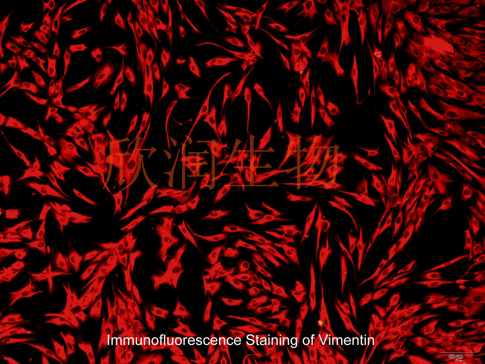
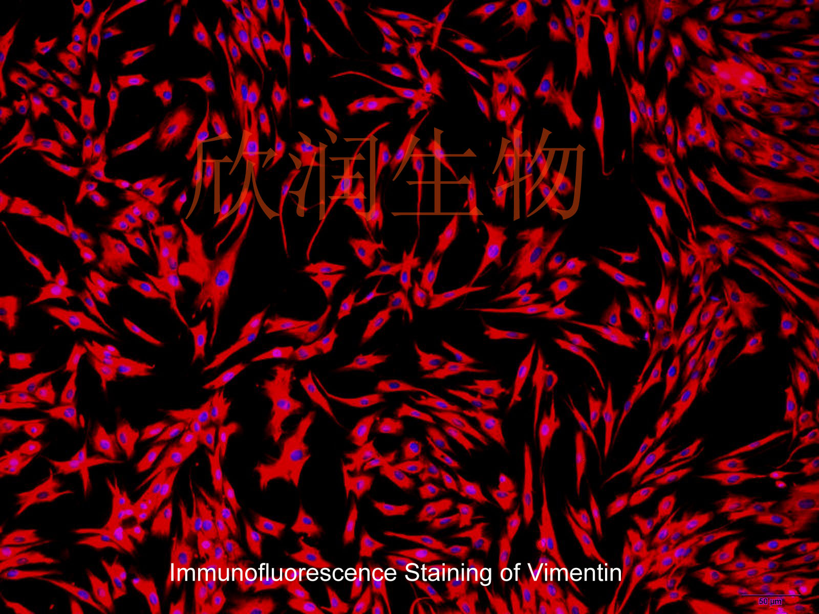
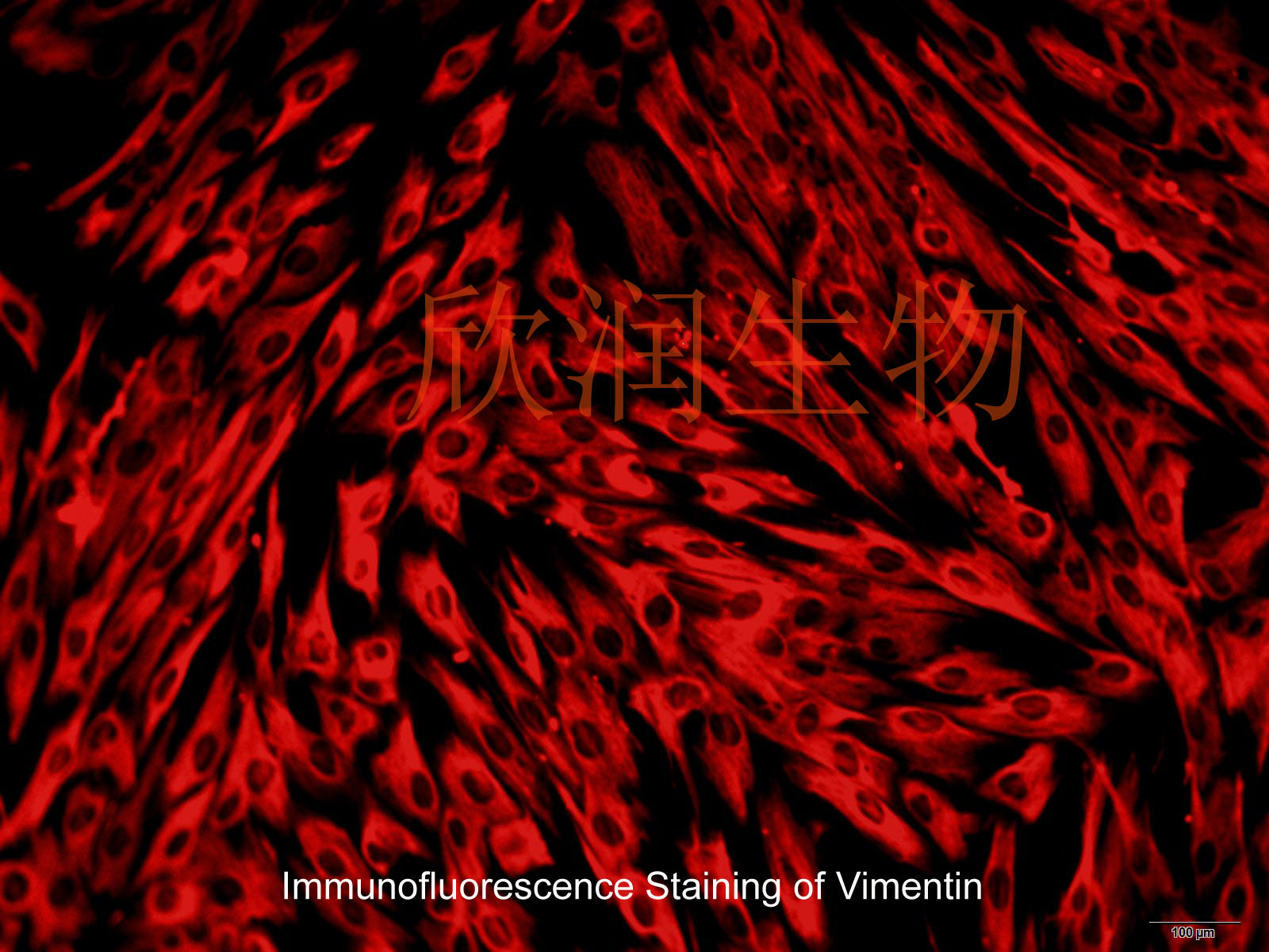
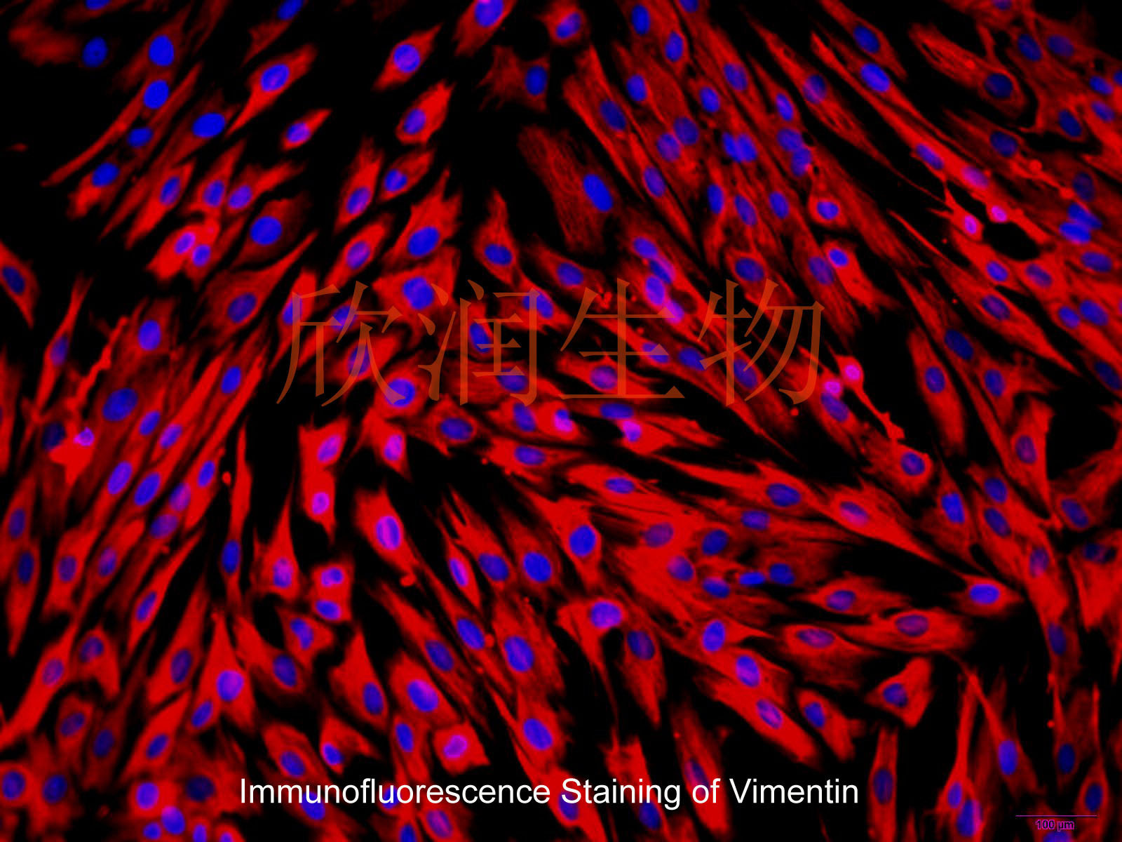
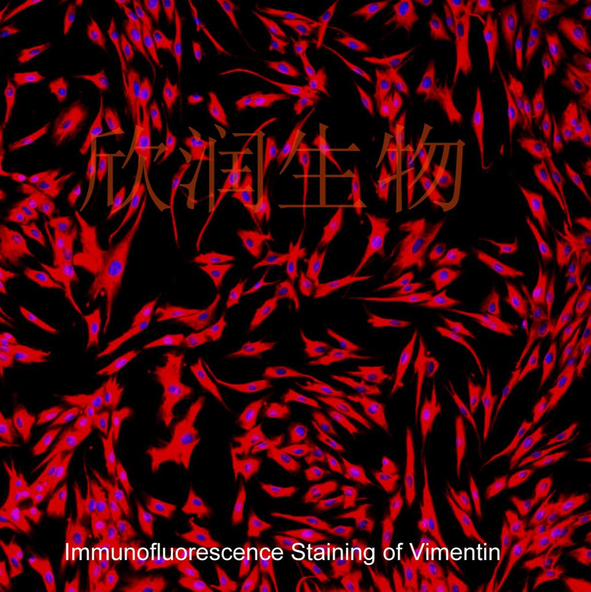
Vimentin免疫荧光检测呈阳性


Telomerase activity in the regenerative basal layer of the epidermis inhuman skin and in immortal and carcinoma-derived skin keratinocytes
Cellular senescence is defined by the limited proliferative capacity of normal cultured cells. Immortal cells overcome this regulation and proliferate indefinitively. One step in the immortalization process may be reactivation of telomerase activity, a ribonucleoprotein complex, which, by de novo synthesized telomeric TTAGGG repeats, can prevent shortening of the telomeres. Here we show that immortal human skin keratinocytes, irrespective of whether they were immortalized by simian virus 40, human papillomavirus 16, or spontaneously, as well as cell lines established from human skin squamous cell carcinomas exhibit telomerase activity. Unexpectedly, four of nine samples of intact human skin also were telomerase positive. By dissecting the skin we could show that the dermis and cultured dermal fibroblasts were telomerase negative. The epidermis and cultured skin keratinocytes, however, reproducibly exhibited enzyme activity. By separating different cell layers of the epidermis this telomerase activity could be assigned to the proliferative basal cells.
风险提示:丁香通仅作为第三方平台,为商家信息发布提供平台空间。用户咨询产品时请注意保护个人信息及财产安全,合理判断,谨慎选购商品,商家和用户对交易行为负责。对于医疗器械类产品,请先查证核实企业经营资质和医疗器械产品注册证情况。
- 作者
- 内容
- 询问日期
 文献和实验
文献和实验Chicken intestinal epithelial cells were obtained from NEWGAINBIO company. Cells were cultured on 37℃, with 5% CO2, in the Ham’s F-12 Nutrient (DMEM/12) that contained the following supplementations: fetal bovine serum (5%), in-sulin (5 µg/mL), transferrin (5 µg/mL), selenium (5 ng/mL), epidermal growth factor (5 ng/mL) and penicillin-streptomycin (100–100 U/mL) for cell culturing (full DMEM/12). Experiments were performed with chicken intestinal epithelial cells and working solutions were prepared with plain DMEM/12 without supplementation. For the investigations, cells were seeded onto 96-well, 24-well or 6-well polystyrene cell culture plates.
Primary hVICs (passage 2) were cultured to 50–60% confluence and infected with pGMLV-SV40T-puro lentivirus (NewgainBio, Wuxi, China) at a multiplicity of infection of 80 supplemented with 5 µg/mL polybrene (Sigma-Aldrich, Buchs, Switzerland).
Tissue was cultured until cells became visible around the tissue, and when the fusion reached 90% (FIGURE 1A) §ask ¦lled with the prepared culturing medium was sent to the company for further immortalisation. Cell immortalisation was done for cell stability and longer-term use. Immortalised cells were cultured with 10% FBS and 1% PS in the DMEM medium. After the cells multiplied and merged, they were routinely passed and grown ( NEWGAINBIO Inc. Wuxi, Jiangsu, China) (FIGURE 1B-C).
Mouse primary cultured renal vascular ECs and VSMCs were obtained from Newgainbio company, which were tested by Factor VIII and α-smooth muscle actin (α-SMA), the marker of ECs and VSMCs. RNeasy Mini Kit was used for RNA extraction, and the above protocols were repeated.
Porcine primary colon epithelial cells (Newgainbio company, Wuxi,China) were cultured in Dulbecco's Modified Eagle's Medium (Solarbio, Beijing, China) containing 10 % fetal bovine serum (BioInd, Kiryat shmona, Lsrael) at 37 ◦C and 5 % CO2 humidity.
实验材料: 1. 正常动物皮肤 2. 不含Ca2+ 和Mg2+ 的1×PBS,添加200000IU/L青霉素、200mg/L链霉素,pH7.2 3. 细胞培养瓶(T25) 4. 手术刀、解剖剪、解剖镊、眼科剪,眼科镊 实验方法: 1. 取正常动物皮肤,置于小培养皿内,用PBS漂洗3-4次,以除去表面血污及毛发; 2. 将洗净的组织用眼科剪剪切成1mm3 左右大小,再用PBS漂洗3次; 3. 用眼科剪将组织块移至培养瓶瓶壁
美少女福音!Science 重磅报道,他们率先实现皮肤无疤痕愈合
增殖的可能。 图片来源:Science 随后,作者也通过标记位于皮肤不同位置的 ENFs,识别出了是位于皮肤深处真皮网状层的 ENFs 被激活产生了 EPFs。 图片来源:Science 在弄清楚 EPFs 的来源后,研究团队探究了是什么导致 ENFs 被激活产生了 EPFs。 由于存在于纤维组织中并负责纤维的改造与产生,成纤维细胞能够感受到纤维组织的力学特性,所以作者认为是受伤后皮肤力学性质的改变,导致了 EPFs 的激活。 通过在不同刚度的模拟组织中培养 ENFs 以及使用抑制剂阻断力学信号
肾细胞CCC-HPF-1 人胚肺二倍体细胞(自建)CCC-ESF-1 人胚胎皮肤成纤维细胞(自建)HFF 人前皮肤成纤维细胞HFSF 人胚胎眼巩膜成纤维细胞HFTF 人胚胎眼Tenon's囊成纤维细胞HK-2 人肾小球上皮细胞HKC 人胚肾上皮细胞HSF 人皮肤成纤维细胞MRC-5 人胚肺成纤维细胞WISH 人羊膜细胞CCC-HEL-1 人胚胎肝正常细胞(自建)CCC-HEK-1 人胚胎肾正常细胞(自建)CCC-HHM-2 人胚胎心肌组织来源细胞(自建)CCC-HPE-2 人胚
 技术资料
技术资料