

- ¥2980
- LMAI Bio
- LM-3245R-FITC
- 中国/美国/欧洲
- 2025年07月07日
- IF=1:50-200
- Human, Mouse, Rat,
- Human, Mouse, Rat,

企业认证
相关产品推荐更多 >


万千商家帮你免费找货
0 人在求购买到急需产品
- 详细信息
- 文献和实验
- 技术资料
- 供应商:
上海联迈生物工程有限公司
- 库存:
大量
- 靶点:
详见说明书
- 级别:
1
- 目录编号:
LM-3245R-FITC
- 克隆性:
多克隆
- 抗原来源:
Rabbit
- 保质期:
1年
- 抗体英文名:
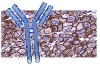
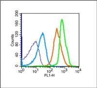
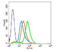
Anti-Phospho-LATS1 (Thr1079)/FITC
- 抗体名:
Anti-Phospho-LATS1 (Thr1079)/FITC
- 标记物:
FITC标记
- 宿主:
Human, Mouse, Rat,
- 适应物种:
Human, Mouse, Rat,
- 免疫原:
详见说明书
- 亚型:
IGg
- 形态:
粉末、液体、冻干粉
- 应用范围:
IF=1:50-200
- 浓度:
1mg/ml
- 保存条件:
-20 °C
- 规格:
100ul
| 英文名称 | Anti-Phospho-LATS1 (Thr1079)/FITC |
| 中文名称 | FITC标记的磷酸化肿瘤抑制基因LATS1抗体 |
| 别 名 | LATS1 (Phospho Thr1079); LATS1 (Phospho T1079); LATS1 (Phospho-Thr1079); Large tumor suppressor homolog 1; Serine threonine protein kinase LATS1; WARTS; WARTS protein kinase; LATS1_HUMAN. |
| 规格价格 | 100ul/2980元 购买 大包装/询价 |
| 说 明 书 | 100ul |
| 产品类型 | 磷酸化抗体 |
| 研究领域 | 肿瘤 免疫学 转录调节因子 |
| 抗体来源 | Rabbit |
| 克隆类型 | Polyclonal |
| 交叉反应 | Human, Mouse, Rat, |
| 产品应用 | IF=1:50-200 not yet tested in other applications. optimal dilutions/concentrations should be determined by the end user. |
| 分 子 量 | 124kDa |
| 性 状 | Lyophilized or Liquid |
| 浓 度 | 1mg/ml |
| 免 疫 原 | KLH conjugated synthesised phosphopeptide derived from human LATS1 around the phosphorylation site of Thr1079 |
| 亚 型 | IgG |
| 纯化方法 | affinity purified by Protein A |
| 储 存 液 | 0.01M TBS(pH7.4) with 1% BSA, 0.03% Proclin300 and 50% Glycerol. |
| 保存条件 | Store at -20 °C for one year. Avoid repeated freeze/thaw cycles. The lyophilized antibody is stable at room temperature for at least one month and for greater than a year when kept at -20°C. When reconstituted in sterile pH 7.4 0.01M PBS or diluent of antibody the antibody is stable for at least two weeks at 2-4 °C. |
| 产品介绍 | background: The protein encoded by this gene is a putative serine/threonine kinase that localizes to the mitotic apparatus and complexes with cell cycle controller CDC2 kinase in early mitosis. The protein is phosphorylated in a cell-cycle dependent manner, with late prophase phosphorylation remaining through metaphase. The N-terminal region of the protein binds CDC2 to form a complex showing reduced H1 histone kinase activity, indicating a role as a negative regulator of CDC2/cyclin A. In addition, the C-terminal kinase domain binds to its own N-terminal region, suggesting potential negative regulation through interference with complex formation via intramolecular binding. Biochemical and genetic data suggest a role as a tumor suppressor. Function: Negative regulator of YAP1 in the Hippo signaling pathway that plays a pivotal role in organ size control and tumor suppression by restricting proliferation and promoting apoptosis. The core of this pathway is composed of a kinase cascade wherein STK3/MST2 and STK4/MST1, in complex with its regulatory protein SAV1, phosphorylates and activates LATS1/2 in complex with its regulatory protein MOB1, which in turn phosphorylates and inactivates YAP1 oncoprotein and WWTR1/TAZ. Phosphorylation of YAP1 by LATS1 inhibits its translocation into the nucleus to regulate cellular genes important for cell proliferation, cell death, and cell migration. Acts as a tumor suppressor which plays a critical role in maintenance of ploidy through its actions in both mitotic progression and the G1 tetraploidy checkpoint. Negatively regulates G2/M transition by down-regulating CDK1 kinase activity. Involved in the control of p53 expression. Affects cytokinesis by regulating actin polymerization through negative modulation of LIMK1. May also play a role in endocrine function. Subunit: Complexes with CDK1 in early mitosis. LATS1-associated CDK1 has no mitotic cyclin partner and no apparent kinase activity. Binds phosphorylated ZYX, locating this protein to the mitotic spindle and suggesting a role for actin regulatory proteins during mitosis. Binds to and colocalizes with LIMK1 at the actomyosin contractile ring during cytokinesis. Interacts (via PPxY motif 2) with YAP1 (via WW domains). Interacts with MOB1A and MOB1B. Interacts with LIMD1, WTIP and AJUBA. Subcellular Location: Cytoplasm, cytoskeleton, centrosome. Note=Localizes to the centrosomes throughout interphase but migrates to the mitotic apparatus, including spindle pole bodies, mitotic spindle, and midbody, during mitosis. Tissue Specificity: Expressed in all adult tissues examined except for lung and kidney. Post-translational modifications: Autophosphorylated and phosphorylated during M-phase of the cell cycle. Phosphorylated by STK3/MST2 at Ser-909 and Thr-1079, which results in its activation. Phosphorylated upon DNA damage, probably by ATM or ATR. Phosphorylation at Ser-464 by NUAK1 and NUAK2 leads to decreased protein level and is required to regulate cellular senescence and cellular ploidy. Similarity: Belongs to the protein kinase superfamily. AGC Ser/Thr protein kinase family. Contains 1 AGC-kinase C-terminal domain. Contains 1 protein kinase domain. Contains 1 UBA domain. Database links: Entrez Gene: 9113 Human Entrez Gene: 16798 Mouse Entrez Gene: 308265 Rat Omim: 603473 Human SwissProt: O95835 Human SwissProt: Q8BYR2 Mouse Unigene: 716697 Human Unigene: 34083 Mouse Unigene: 29152 Rat Important Note: This product as supplied is intended for research use only, not for use in human, therapeutic or diagnostic applications. |
风险提示:丁香通仅作为第三方平台,为商家信息发布提供平台空间。用户咨询产品时请注意保护个人信息及财产安全,合理判断,谨慎选购商品,商家和用户对交易行为负责。对于医疗器械类产品,请先查证核实企业经营资质和医疗器械产品注册证情况。
 文献和实验
文献和实验3 篇 Nature 连发,解决世纪难题,周期蛋白的毁灭调控蕴含癌症患者的新生
在 cyclin D 上,被泛素化标记的 cyclin D 将会被蛋白酶体降解。此外研究还发现,AMBRA1 缺失会导致 cyclin D 和 MYC 蛋白水平升高。cyclin D 与 CDK4/6 结合,使 RB1 蛋白磷酸化。磷酸化 RB1 释放 E2F 转录因子来驱动细胞周期进程所需基因的表达。在 AMBRA1 缺失的细胞中,cyclin D 也能与 CDK2 激酶形成复合物,使癌细胞能够抵抗 CDK4/6 抑制剂的治疗。高水平的 cyclin D 会促进细胞增殖,从而导致 DNA 损伤、复制应激
去乙酰化酶 (HDAC) 酶是共抑制因子,通过降低赖氨酸乙酰化水平和增加染色体凝聚而逆转乙酰化的作用。Sirtuins(沉默信息调节因子)是一组 NAD 依赖性去乙酰化酶,靶向组蛋白。顾名思义,它们通过低乙酰化组蛋白维持基因沉默,据报道有助于维持基因组稳定性。而乙酰化最早是在组蛋白中检测到的,胞浆蛋白也被报道是乙酰化的,因此乙酰化似乎在细胞生物学中发挥着比单纯的转录调控更大的作用。此外,乙酰化修饰和其他翻译后修饰,包括磷酸化、泛素化和甲基化之间的串扰,可以改变乙酰化蛋白的生物学功能。可通过使用乙酰赖氨酸特异性抗体
Cell Metab:浙大吕志民团队揭示肿瘤细胞 Warburg 效应促进肿瘤免疫逃逸
2 从线粒体外膜脱落,进入细胞浆中。在细胞浆中,HK2 结合 NF-κB 的抑制因子 IκBα。 重要的是,HK2 发挥了不依赖于其经典代谢功能的新功能,即作为一个蛋白激酶磷酸化 IκBα 的 Thr 291 位点。该磷酸化促进了蛋白酶 μ-calpain 与 IκBα 的结合并进一步降解 IκBα,进而使转录因子 NF-κB 入核,最终促进了 PD-L1 的表达并导致了肿瘤的免疫逃逸。作者还发现,使用己糖激酶的抑制剂与 PD-1 抗体联用治疗小鼠胶质瘤,可以显著提升 PD-1 抗体的治疗
 技术资料
技术资料暂无技术资料 索取技术资料

