

- ¥2980
- LMAI Bio
- LM-5351R-FITC
- 中国/美国/欧洲
- 2026年05月21日
- IF=1:50-200
- Human, Mouse, Rat, Dog, Horse,
- Human, Mouse, Rat, Dog, Horse,

企业认证
相关产品推荐更多 >


万千商家帮你免费找货
0 人在求购买到急需产品
- 详细信息
- 文献和实验
- 技术资料
- 供应商:
上海联迈生物工程有限公司
- 库存:
大量
- 靶点:
详见说明书
- 级别:
1
- 目录编号:
LM-5351R-FITC
- 克隆性:
多克隆
- 抗原来源:
Rabbit
- 保质期:
1年
- 抗体英文名:
Anti-phospho-Filamin A(Ser2522)/FITC
- 抗体名:
Anti-phospho-Filamin A(Ser2522)/FITC
- 标记物:
FITC标记
- 宿主:
Human, Mouse, Rat, Dog, Horse,
- 适应物种:
Human, Mouse, Rat, Dog, Horse,
- 免疫原:
详见说明书
- 亚型:
IGg
- 形态:
粉末、液体、冻干粉
- 应用范围:
IF=1:50-200
- 浓度:
1mg/ml
- 保存条件:
-20 °C
- 规格:
100ul
| 英文名称 | Anti-phospho-Filamin A(Ser2522)/FITC |
| 中文名称 | FITC标记的磷酸化细丝蛋白A抗体 |
| 别 名 | ABP 280; ABP 280 like protein; ABP-280; ABP280A; ABPA antibody Actin binding like protein; Actin binding protein 280; Actin-binding protein 280; Alpha filamin; Alpha-filamin; APBX; cb967; Dilp2; Endothelial actin binding protein; Endothelial actin-binding protein; Filamin 1; Filamin A alpha actin binding protein 280; Filamin A; Filamin-1; Filamin-A; FLN; FLN-A; FLN1; FLNA; FLNA_HUMAN; FMD; MNS; NHBP; Non muscle filamin; Non-muscle filamin; OPD; OPD1; OPD2. |
| 规格价格 | 100ul/2980元 购买 大包装/询价 |
| 说 明 书 | 100ul |
| 产品类型 | 磷酸化抗体 |
| 研究领域 | 免疫学 神经生物学 通道蛋白 结合蛋白 |
| 抗体来源 | Rabbit |
| 克隆类型 | Polyclonal |
| 交叉反应 | Human, Mouse, Rat, Dog, Horse, |
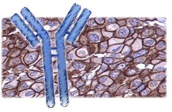
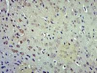
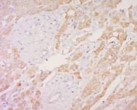
| 产品应用 | IF=1:50-200 not yet tested in other applications. optimal dilutions/concentrations should be determined by the end user. |
| 分 子 量 | 291kDa |
| 性 状 | Lyophilized or Liquid |
| 浓 度 | 1mg/ml |
| 免 疫 原 | KLH conjugated Synthesised phosphopeptide derived from human FLNA around the phosphorylation site of Ser2522 |
| 亚 型 | IgG |
| 纯化方法 | affinity purified by Protein A |
| 储 存 液 | 0.01M TBS(pH7.4) with 1% BSA, 0.03% Proclin300 and 50% Glycerol. |
| 保存条件 | Store at -20 °C for one year. Avoid repeated freeze/thaw cycles. The lyophilized antibody is stable at room temperature for at least one month and for greater than a year when kept at -20°C. When reconstituted in sterile pH 7.4 0.01M PBS or diluent of antibody the antibody is stable for at least two weeks at 2-4 °C. |
| 产品介绍 | background: The protein encoded by this gene is an actin-binding protein that crosslinks actin filaments and links actin filaments to membrane glycoproteins. The encoded protein is involved in remodeling the cytoskeleton to effect changes in cell shape and migration. This protein interacts with integrins, transmembrane receptor complexes, and second messengers. Defects in this gene are a cause of several syndromes, including periventricular nodular heterotopias (PVNH1, PVNH4), otopalatodigital syndromes (OPD1, OPD2), frontometaphyseal dysplasia (FMD), Melnick-Needles syndrome (MNS), and X-linked congenital idiopathic intestinal pseudoobstruction (CIIPX). Two transcript variants encoding different isoforms have been found for this gene.[provided by RefSeq, Mar 2009] Function: Promotes orthogonal branching of actin filaments and links actin filaments to membrane glycoproteins. Anchors various transmembrane proteins to the actin cytoskeleton and serves as a scaffold for a wide range of cytoplasmic signaling proteins. Interaction with FLNA may allow neuroblast migration from the ventricular zone into the cortical plate. Tethers cell surface-localized furin, modulates its rate of internalization and directs its intracellular trafficking. Involved in ciliogenesis. Subunit: Interacts with PDLIM2 (By similarity). Homodimer. Interacts with FCGR1A, FLNB, FURIN, HSPB7, INPPL1, KCND2, MYOT, MYOZ1, ARHGAP24, PSEN1, PSEN2 and ECSCR. Interacts also with various other binding partners in addition to filamentous actin. Interacts (via N-terminus) with MIS18BP1 (via N-terminus). Interacts (via N-terminus) with TAF1B. Interacts with TMEM67 (via C-terminus) and MKS1. Subcellular Location: Cytoplasm, cell cortex. Cytoplasm, cytoskeleton. Tissue Specificity: Ubiquitous. Post-translational modifications: Phosphorylated upon DNA damage, probably by ATM or ATR. Phosphorylation extent changes in response to cell activation. DISEASE: Defects in FLNA are the cause of periventricular nodular heterotopia type 1 (PVNH1) [MIM:300049]; also called nodular heterotopia, bilateral periventricular (NHBP or BPNH). PVNH is a developmental disorder characterized by the presence of periventricular nodules of cerebral gray matter, resulting from a failure of neurons to migrate normally from the lateral ventricular proliferative zone, where they are formed, to the cerebral cortex. PVNH1 is an X-linked dominant form. Heterozygous females have normal intelligence but suffer from seizures and various manifestations outside the central nervous system, especially related to the vascular system. Hemizygous affected males die in the prenatal or perinatal period. Defects in FLNA are the cause of periventricular nodular heterotopia type 4 (PVNH4) [MIM:300537]; also known as periventricular heterotopia Ehlers-Danlos variant. PVNH4 is characterized by nodular brain heterotopia, joint hypermobility and development of aortic dilation in early adulthood. Defects in FLNA are the cause of otopalatodigital syndrome type 1 (OPD1) [MIM:311300]. OPD1 is an X-linked dominant multiple congenital anomalies disease mainly characterized by a generalized skeletal dysplasia, mild mental retardation, hearing loss, cleft palate, and typical facial anomalies. OPD1 belongs to a group of X-linked skeletal dysplasias known as oto-palato-digital syndrome spectrum disorders that also include OPD2, Melnick-Needles syndrome (MNS), and frontometaphyseal dysplasia (FMD). Remodeling of the cytoskeleton is central to the modulation of cell shape and migration. FLNA is a widely expressed protein that regulates re-organization of the actin cytoskeleton by interacting with integrins, transmembrane receptor complexes and second messengers. Males with OPD1 have cleft palate, malformations of the ossicles causing deafness and milder bone and limb defects than those associated with OPD2. Obligate female carriers of mutations causing both OPD1 and OPD2 have variable (often milder) expression of a similar phenotypic spectrum. Defects in FLNA are the cause of otopalatodigital syndrome type 2 (OPD2) [MIM:304120]; also known as cranioorodigital syndrome. OPD2 is a congenital bone disorder that is characterized by abnormally modeled, bowed bones, small or absent first digits and, more variably, cleft palate, posterior fossa brain anomalies, omphalocele and cardiac defects. Defects in FLNA are the cause of frontometaphyseal dysplasia (FMD) [MIM:305620]. FMD is a congenital bone disease characterized by supraorbital hyperostosis, deafness and digital anomalies. Defects in FLNA are the cause of Melnick-Needles syndrome (MNS) [MIM:309350]. MNS is a severe congenital bone disorder characterized by typical facies (exophthalmos, full cheeks, micrognathia and malalignment of teeth), flaring of the metaphyses of long bones, s-like curvature of bones of legs, irregular constrictions in the ribs, and sclerosis of base of skull. Defects in FLNA are the cause of X-linked congenital idiopathic intestinal pseudoobstruction (CIIPX) [MIM:300048]. CIIPX is characterized by a severe abnormality of gastrointestinal motility due to primary qualitative defects of enteric ganglia and nerve fibers. Affected individuals manifest recurrent signs of intestinal obstruction in the absence of any mechanical lesion. Defects in FLNA are the cause of FG syndrome type 2 (FGS2) [MIM:300321]. FG syndrome (FGS) is an X-linked disorder characterized by mental retardation, relative macrocephaly, hypotonia and constipation. Defects in FLNA are the cause of terminal osseous dysplasia (TOD) [MIM:300244]. A rare X-linked dominant male-lethal disease characterized by skeletal dysplasia of the limbs, pigmentary defects of the skin and recurrent digital fibroma during infancy. A significant phenotypic variability is observed in affected females. Defects in FLNA are the cause of cardiac valvular dysplasia X-linked (CVDX) [MIM:314400]. A rare X-linked heart disease characterized by mitral and/or aortic valve regurgitation. The histologic features include fragmentation of collagenous bundles within the valve fibrosa and accumulation of proteoglycans, which produces excessive valve tissue leading to billowing of the valve leaflets. Note=Defects in FLNA may be a cause of macrothrombocytopenia, a disorder characterized by subnormal levels of blood platelets. Blood platelets are abonormally enlarged. Similarity: Belongs to the filamin family. Contains 1 actin-binding domain. Contains 2 CH (calponin-homology) domains. Contains 24 filamin repeats. Database links: Entrez Gene: 2316 Human Entrez Gene: 192176 Mouse Entrez Gene: 293860 Rat Omim: 300017 Human SwissProt: P21333 Human SwissProt: Q8BTM8 Mouse Unigene: 195464 Human Unigene: 295533 Mouse Unigene: 4213 Rat Important Note: This product as supplied is intended for research use only, not for use in human, therapeutic or diagnostic applications. 内皮细胞肌动蛋白结合蛋白(肌动结合蛋白样蛋白) |
风险提示:丁香通仅作为第三方平台,为商家信息发布提供平台空间。用户咨询产品时请注意保护个人信息及财产安全,合理判断,谨慎选购商品,商家和用户对交易行为负责。对于医疗器械类产品,请先查证核实企业经营资质和医疗器械产品注册证情况。
 文献和实验
文献和实验双重染色法免疫荧光组织化学染色步骤若在同一标本中有A和B两种抗原需要同时显示,A抗原的抗体用FITC标记,B抗原的抗体用TRITC标记,可采用以下染色方法:免疫荧光细胞化学双重染鱼法原代培养胚胎14天大鼠端脑细胞:在骨发生形态蛋白―{诱导下,乙酰胆碱转移酶(呈绿色荧光)和同源域蛋白Islet―1(呈红色荧光)共存于细胞质内(激光扫描共聚焦显微镜观察)1.一步双染色法先将两种荧光标{己抗体按适当比例混合(A+B),按直接法进行染色。2.二步双染色法先用TRITC标记的A抗体进行免疫荧光染色,再用
染色,不必洗去,再用FITC标记的A抗体染色,按间接法进行。结果:A抗原阳性荧光呈现绿色,B抗原阳性呈现桔红色荧光。3、荧光抗体再染色法若切片或其他标本经某种荧光抗体染色后,未获得阳性结果,而又疑有另外的病原体存在时,可用相应的荧光抗体再染色。有时存档蜡块不能再用以切片,也可用存档的HE染色标本,褪去盖片和颜色,再作免疫荧光或其它免疫细胞化学的染色。二、荧光抗原染色法某些抗原可以用荧光素标记,制成荧光抗原,标记荧光素的方法与制备荧光抗体方法相同。用荧光抗原可以直接检查细胞或组织内的相应抗体,特异性较好
免疫荧光单标记和双标记的方法 1免疫荧光单标记方法 免疫 荧光单标记是指只标记一种蛋白质分子,方法比较简单,只要按照染色步骤去做,通常不存在太多的问题。但要注意固定液的选择,固定液选择的合适与否,可能会直接影响染色结果。具体染色方法如下。 1)所需材料与试剂 (1)培养在盖玻片或玻璃培养皿中融合程度达到60%一70%的细胞。 (2)一抗、FITC或TRITC标记的二抗。 (3)4%多聚甲醛固定液或冷
 技术资料
技术资料暂无技术资料 索取技术资料

