


- ¥2219
- AAT
- 22658
- USA
- 2026年01月17日

企业认证
相关产品推荐更多 >


万千商家帮你免费找货
0 人在求购买到急需产品
- 详细信息
- 询价记录
- 文献和实验
- 技术资料
- 英文名:
Cell Navigator™ 溶酶体染色试剂盒 *红色荧光*
- 保存条件:
低温
- 规格:
500 Tests
Cell Navigator™ 溶酶体染色试剂盒 *红色荧光*
| Catalog | Unit Size | Price(USD) | Additional Information |
| 22655 | 500 Tests | ¥2219 | Blue Fluorescence |
| 22659 | 500 Tests | ¥2219 | Deep Red Fluorescence |
| 22656 | 500 Tests | ¥2219 | Green Fluorescence |
| 22651 | 500 Tests | ¥2973 | Green Fluorescence with 405 nm Excitation |
| 22652 | 500 Tests | ¥2973 | NIR Fluorescence |
| 22657 | 500 Tests | ¥2219 | Orange Fluorescence |
| 22658 | 500 Tests | ¥2219 | Red Fluorescence |
Platform
| Fluorescence microscope | |
| Excitation | TRITC filter |
| Emission | TRITC filter |
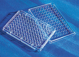
| Recommended plate | Black wall/clear bottom |
Components
| Component A: LysoBrite™ Red | 1 vial (100 µL, 500X DMSO stock solution) |
| Component B: Live Cell Staining Buffer | 1 bottle (50 mL) |
Example protocol
At a glance
Protocol summary
- Prepare cells
- Add LysoBrite™ Red working solution
- Incubate at 37°C for 30 minutes
- Wash the cells
- Analyze the cells under fluorescence microscope at Ex/Em = 575/600 nm (TRITC filter set)
Important notes
Thaw all the kit components at room temperature before starting the experiment.
Preparation of cell samples
Preparation of working solution
Add 20 µL of 500X LysoBrite™ Red (Component A) to 10 mL of Live Cell Staining Buffer (Component B) to make LysoBrite™ Red working solution. Protect from light. Note: 20 µL of 500X LysoBrite™ Red (Component A) is enough for one 96-well plate. The optimal concentration of the fluorescent lysosome indicator varies depending on the specific application. The staining conditions may be modified according to the particular cell type and the permeability of the cells or tissues to the probe.
Procedure
For adherent cells:
- Grow cells either in a 96-well black wall/clear bottom plate (100 µL/well/96-well plate) or on cover-slips inside a petri dish filled with the appropriate culture medium.
- When cells reach the desired confluence, add equal volume of LysoBrite™ Red working solution.
- Incubate the cells in a 37°C, 5% CO2 incubator for 30 minutes.
- Wash the cells twice with pre-warmed (37°C) Hanks and 20 mM Hepes buffer (HBSS) or buffer of your choice, fill the cell wells with HBSS or growth medium.
- Observe the cells using a fluorescence microscope with TRITC filter set (Ex/Em = 575/600 nm). Note: It is recommended to increase either the labeling concentration or the incubation time to allow the dye to accumulate if the cells do not appear to be sufficiently stained.
For suspension cells:
- Add equal volume of LysoBrite™ Red working solution into the cells.
- Incubate the cells in a 37°C, 5% CO2 incubator for 30 minutes.
- Wash the cells twice with pre-warmed (37°C) Hanks and 20 mM Hepes buffer (HBSS) or buffer of your choice, fill the cell wells with HBSS or growth medium.
- Observe the cells using a fluorescence microscope with TRITC filter set (Ex/Em = 575/600 nm). Note: It is recommended to increase either the labeling concentration or the incubation time to allow the dye to accumulate if the cells do not appear to be sufficiently stained. Suspension cells may be attached to cover-slips that have been treated with BD Cell-Tak® (BD Biosciences) and stained as adherent cells.
风险提示:丁香通仅作为第三方平台,为商家信息发布提供平台空间。用户咨询产品时请注意保护个人信息及财产安全,合理判断,谨慎选购商品,商家和用户对交易行为负责。对于医疗器械类产品,请先查证核实企业经营资质和医疗器械产品注册证情况。
- 作者
- 内容
- 询问日期
 文献和实验
文献和实验韶光逐浅 我想问一下Hoechst凋亡染色试剂盒国产的哪个公司的好啊? yunitongxing 原装进口试剂 韶光逐浅 好的,多谢啦 本文由丁香园论坛提供,想了解更多有用的、有意思的前沿资讯以及酷炫的实验方法的你,都可以成为师兄的好伙伴 师兄微信号:shixiongcoming
案例一:DAB染色后切片着色一片黄/背景深 背景:奥运会期间我们课题组研究生都在实验室做实验,许多从北京购买的试剂买不到,对我们的许多实验产生了很大的影响。 我以前一直用中杉金桥的Sp三步法染色试剂盒,效果不错,在奥运会期间我准备做同批实验分不同批次酶免疫组化实验,第一批组化实验结果很好,抗体浓度感觉比较合适(Santa Cruz公司1:200),等做第二批时发现封闭血清不够了,为了保证实验顺利进行,我又从福州迈新顶了一个SP试剂盒,同时抗体稀释液也不够了,我又重新从博士
* 植物样本突破:植物脂质在信号转导、抗逆性中至关重要。传统方法需数百克起始材料、数天时间;Invent柱式法仅需毫克级样品、1小时内即可完成,彻底解决植物亚细胞脂质组学的样品制备难题。 Minute™植物质膜分离试剂盒(Cat# SM-005-P) Minute™植物脂筏分离试剂盒(Cat# PL-051) Minute™植物内质网分离试剂盒(Cat# PR-048) Minute™植物高尔基体分离试剂盒(Cat# PG-049) 此外,Invent 技术覆盖线粒体、溶酶体、内体、外泌体等全套亚
 技术资料
技术资料暂无技术资料 索取技术资料

