

- ¥780 - 2200
- LMAI Bio
- LM-4770R
- 进口/国产
- 2026年05月13日
- WB=1:500-2000 ELISA=1:500-1000 Flow-Cyt=1μg/Test (石蜡切片需做抗原修复)
- Rabbit
- Human, Mouse, Rat,

企业认证
相关产品推荐更多 >
万千商家帮你免费找货
0 人在求购买到急需产品
- 详细信息
- 文献和实验
- 技术资料
- 供应商:
上海联迈生物工程有限公司
- 库存:
大量
- 目录编号:
LM-4770R
- 克隆性:
多克隆
- 抗原来源:
Rabbit
- 保质期:
1年
- 抗体英文名:
CD133
- 抗体名:
造血干细胞抗原CD133抗体
- 宿主:
Rabbit
- 适应物种:
Human, Mouse, Rat,
- 免疫原:
KLH conjugated synthetic peptide derived from human CD133:508-552/865 <Extracellular>
- 亚型:
IgG
- 形态:
Lyophilized or Liquid
- 应用范围:
WB=1:500-2000 ELISA=1:500-1000 Flow-Cyt=1μg/Test (石蜡切片需做抗原修复)
- 浓度:
1mg/ml
- 保存条件:
Store at -20 °C
- 规格:
50ul 100ul 200ul
| 英文名称 | CD133 |
| 中文名称 | 造血干细胞抗原CD133抗体 |
| 别 名 | AC133; Antigen AC133; Hematopoietic stem cell antigen; hProminin; PROM1; Prominin I; Prominin 1; Prominin1; Prominin-1; Prominin like protein 1 precursor; Prominin mouse like 1; prominin1; PROML1; CD133; CORD12; MCDR2; MSTP061; PROML1; RP41; STGD4. |
| 规格价格 | 50ul/780元 购买 100ul/1380元 购买 200ul/2200元 购买 大包装/询价 |
| 说 明 书 | 50ul 100ul 200ul |
| 研究领域 | 肿瘤 细胞生物 免疫学 干细胞 细胞类型标志物 |
| 抗体来源 | Rabbit |
| 克隆类型 | Polyclonal |
| 交叉反应 | Human, Mouse, Rat, |
| 产品应用 | WB=1:500-2000 ELISA=1:500-1000 Flow-Cyt=1μg/Test (石蜡切片需做抗原修复) not yet tested in other applications. optimal dilutions/concentrations should be determined by the end user. |
| 分 子 量 | 95kDa |
| 细胞定位 | 细胞膜 |
| 性 状 | Lyophilized or Liquid |
| 浓 度 | 1mg/ml |
| 免 疫 原 | KLH conjugated synthetic peptide derived from human CD133:508-552/865 <Extracellular> |
| 亚 型 | IgG |
| 纯化方法 | affinity purified by Protein A |
| 储 存 液 | 0.01M TBS(pH7.4) with 1% BSA, 0.03% Proclin300 and 50% Glycerol. |
| 保存条件 | Store at -20 °C for one year. Avoid repeated freeze/thaw cycles. The lyophilized antibody is stable at room temperature for at least one month and for greater than a year when kept at -20°C. When reconstituted in sterile pH 7.4 0.01M PBS or diluent of antibody the antibody is stable for at least two weeks at 2-4 °C. |
| PubMed | PubMed |
| 产品介绍 | background: This gene encodes a pentaspan transmembrane glycoprotein. The protein localizes to membrane protrusions and is often expressed on adult stem cells, where it is thought to function in maintaining stem cell properties by suppressing differentiation. Mutations in this gene have been shown to result in retinitis pigmentosa and Stargardt disease. Expression of this gene is also associated with several types of cancer. This gene is expressed from at least five alternative promoters that are expressed in a tissue-dependent manner. Multiple transcript variants encoding different isoforms have been found for this gene. [provided by RefSeq, Mar 2009] Function: Binds cholesterol in cholesterol-containing plasma membrane microdomains. Proposed to play a role in apical plasma membrane organization of epithelial cells. During early retinal development acts as a key regulator of disk morphogenesis. Involved in regulation of MAPK and Akt signaling pathways. In neuroblastoma cells suppresses cell differentiation such as neurite outgrowth in a RET-dependent manner. Subunit: Interacts with CDHR1 and with actin filaments. Subcellular Location: Cell projection, cilium, photoreceptor outer segment. Isoform 1: Apical cell membrane; Multi-pass membrane protein. Cell projection, microvillus membrane; Multi-pass membrane protein. Note=Found in extracellular membrane particles in various body fluids such as cerebrospinal fluid, saliva, seminal fluid and urine. Tissue Specificity: Isoform 1 is selectively expressed on CD34 hematopoietic stem and progenitor cells in adult and fetal bone marrow, fetal liver, cord blood and adult peripheral blood. Isoform 1 is not detected on other blood cells. Isoform 1 is also expressed in a number of non-lymphoid tissues including retina, pancreas, placenta, kidney, liver, lung, brain and heart. Found in saliva within small membrane particles. Isoform 2 is predominantly expressed in fetal liver, skeletal muscle, kidney, and heart as well as adult pancreas, kidney, liver, lung, and placenta. Isoform 2 is highly expressed in fetal liver, low in bone marrow, and barely detectable in peripheral blood. Isoform 2 is expressed on hematopoietic stem cells and in epidermal basal cells (at protein level). Expressed in adult retina by rod and cone photoreceptor cells (at protein level). Post-translational modifications: Isoform 1 and isoform 2 are glycosylated. DISEASE: Defects in PROM1 are the cause of retinitis pigmentosa type 41 (RP41) [MIM:612095]; also known as retinal degeneration autosomal recessive prominin-related. RP is a retinal dystrophy belonging to the group of pigmentary retinopathies. RP is characterized by retinal pigment deposits visible on fundus examination and primary loss of rod photoreceptor cells followed by secondary loss of cone photoreceptors. Patients typically have night vision blindness and loss of midperipheral visual field. As their condition progresses, they lose their far peripheral visual field and eventually central vision as well. Defects in PROM1 are the cause of cone-rod dystrophy type 12 (CORD12) [MIM:612657]. CORD12 is an inherited retinal dystrophy characterized by retinal pigment deposits visible on fundus examination, predominantly in the macular region, and initial loss of cone photoreceptors followed by rod degeneration. This leads to decreased visual acuity and sensitivity in the central visual field, followed by loss of peripheral vision. Severe loss of vision occurs earlier than in retinitis pigmentosa. Defects in PROM1 are the cause of Stargardt disease type 4 (STGD4) [MIM:603786]. Stargardt disease is the most common hereditary macular degeneration. It is characterized by decreased central vision, atrophy of the macula and underlying retinal pigment epithelium, and frequent presence of prominent flecks in the posterior pole of the retina. Defects in PROM1 are the cause of retinal macular dystrophy type 2 (MCDR2) [MIM:608051]. MCDR2 is a bull's-eye macular dystrophy characterized by bilateral annular atrophy of retinal pigment epithelium at the macula. Similarity: Belongs to the prominin family. SWISS: O43490 Gene ID: 8842 Database links: Entrez Gene: 8842 Human Entrez Gene: 19126 Mouse Entrez Gene: 60357 Rat Omim: 604365 Human SwissProt: O43490 Human SwissProt: O54990 Mouse Unigene: 614734 Human Unigene: 6250 Mouse Unigene: 144589 Rat Important Note: This product as supplied is intended for research use only, not for use in human, therapeutic or diagnostic applications. 一般认为,VEGFR2(血管内皮生长因子受体2)是HSCs(造血干细胞)的特异性的表面标志。近来经研究发现CD133分子是HSCs(造血干细胞)特异性标志。CD133即AC133,是一个新发现的HSCs(造血干细胞)表面标志,在HSCs(造血干细胞)分化成熟过程中,CD133的含量迅速降低。EPCs(血管内皮前体细胞)区别于成熟内皮细胞的主要标志是CD133。 经研究发现内皮细胞不能结合CD133的抗体。证实分化成熟的内皮细胞不具有CD133。这些说明CD133可以作为EPCs(血管内皮前体细胞)区别于成熟内皮细胞的一个表面标志. |
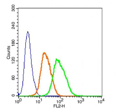
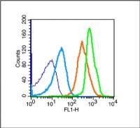
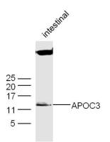
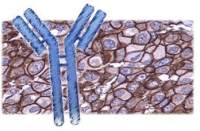
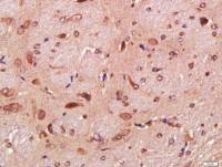
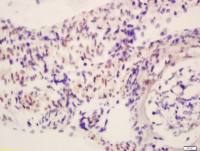
| 产品图片 | 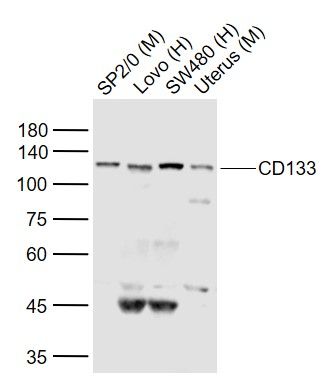 Sample: Lovo Cell (Human) Lysate at 30 ug Primary: Anti-CD133 (bs-4770R)at 1/300 dilution Secondary: IRDye800CW Goat Anti-Rabbit IgG at 1/20000 dilution Predicted band size: 95kD Observed band size: 120kD 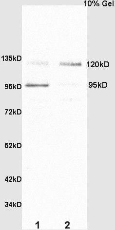 Sample: SP2/0 Cell (Mouse) Lysate at 40 ug Colon carcinoma (Human) Lysate at 40 ug Primary: Anti-CD133 (bs-4770R) at 1/300 dilution Secondary: HRP conjugated Goat-Anti-rabbit IgG (bs-0295G-HRP) at 1/5000 dilution Predicted band size: 95 kD Observed band size: 95/120 kD 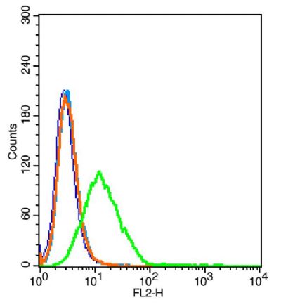 Blank control: 293T(blue). Primary Antibody:Rabbit Anti-CD133 antibody(bs-4770R), Dilution: 5μg in 100 1μL 1X PBS containing 0.5% BSA; Isotype Control Antibody: Rabbit IgG(orange) ,used under the same conditions ); Secondary Antibody: Goat anti-rabbit IgG-PE(white blue), Dilution: 1:200 in 1 X PBS containing 0.5% BSA.  The blue histogram is unstained cells(HepG 2). The Orange histogram is cells stained with Rabbit IgG/FITC (bs-0295P-FITC) The green histogram is cells stained with Rabbit Anti-CD133/FITC Conjugated antibody (bs-4770R-FITC). Isotype control: Cell lines treated with Rabbit IgG/FITC (bs-0295P-FITC) instead of the primary antibody to confirm that primary antibody binding is 2μg/5μg/1μg in 100μL 1 X PBS containing 0.5% BSA.  The blue histogram is unstained cells(HepG 2). The Orange histogram is cells stained with Rabbit IgG/PE (bs-0295P-PE) The green histogram is cells stained with Rabbit Anti-CD133/PE Conjugated antibody (bs-4770R-PE). Isotype control: Cell lines treated with Rabbit IgG/PE (bs-0295P-PE) instead of the primary antibody to confirm that primary antibody binding is specific. 2μg/5μg/10μg in 100μL 1 X PBS containing 0.5% BSA. |
风险提示:丁香通仅作为第三方平台,为商家信息发布提供平台空间。用户咨询产品时请注意保护个人信息及财产安全,合理判断,谨慎选购商品,商家和用户对交易行为负责。对于医疗器械类产品,请先查证核实企业经营资质和医疗器械产品注册证情况。
 文献和实验
文献和实验MACSQuant Tyto 分选有效提高造血干细胞移植与基因修饰
由于基因编辑技术的日益演进,不仅细胞核内基因编辑可修复 DNA 缺陷帮助治疗遗传疾病 (如脊髓性肌萎缩症),也可导入特定基因增强细胞功能消灭恶性肿瘤(如 CART 治疗 B 细胞淋巴瘤)。今年 7 月 Nature 发表的线粒体基因编辑工具更是横空出世,这对于 mtDNA 突变引起的母系遗传 Leigh 综合征、线粒体肌病等线粒体遗传疾病的研究、治疗和治愈带来了希望。 如今,结合基因治疗的造血干细胞移植,已有多项重大研究与临床应用正如火如荼地展开。如 2019 年欧盟批准基因治疗与干细
异构体AC133-2, 最近已经被克隆并鉴定为可被AC133抗体识别的原始表面抗原。CD133可以作为用CD34筛选HSC和体外扩增的补充。CD133+富集的亚类可以以同CD34+ 富集的亚类扩增的方式扩增,从而可保留多系增殖的能力。最近的研究为CD133的表达不限于原始血细胞提供了证据,同时也确定了非造血组织中一类独特的细胞群体。来源于外周血的CD133+ 可被体外诱导分化为内皮细胞。并且,can be induced to differentiate into endothelial
到经多聚赖氨酸处理的玻片上,56℃温箱中烘干,PBS 洗3 次,4% 多聚甲醛室温固定20min,PBS 洗2 次,0.1% Triton-X100 孵育10min,PBS 洗2 次;1%BSA封闭,室温孵育1h,PBS 洗2 次;分别加入适量以封闭液稀释的第一抗体CD133、Nestin,4℃孵育过夜;PBS 洗3 次,加入FITC(两种抗体只能加FITC,因为转染后的干细胞表达红色荧光蛋白)耦联的二抗,室温避光孵育2h ;PBS 洗3 次,以50%、70%、95% 和100% 四个浓度梯度的乙醇
 技术资料
技术资料暂无技术资料 索取技术资料

