

- ¥1580
- 康朗生物
- kl-5355R
- 中国/美国/德国
- 2026年01月04日
- WB=1:500-2000 ELISA=1:500-1000 IHC-P=1:400-800 IHC-F=1:400-800 Flow-Cyt=3μg/Test ICC=1:100-500 IF=1:100-500
- Rabbit
- Human, Mouse, Rat, Pig,

企业认证
相关产品推荐更多 >


万千商家帮你免费找货
0 人在求购买到急需产品
- 详细信息
- 文献和实验
- 技术资料
- 供应商:
上海康朗生物科技有限公司
- 库存:
大量
- 目录编号:
kl-5355R
- 克隆性:
多克隆
- 抗原来源:
Rabbit
- 保质期:
12个月
- 抗体英文名:
phospho-GFAP (Ser8) antibody
- 抗体名:
磷酸化胶质纤维酸性蛋白抗体
- 宿主:
Rabbit
- 适应物种:
Human, Mouse, Rat, Pig,
- 免疫原:
KLH conjugated Synthesised phosphopeptide derived from human GFAP around the phosphorylation site of Ser8:IT(p-S)A
- 亚型:
IgG
- 形态:
冻干粉或液体
- 应用范围:
WB=1:500-2000 ELISA=1:500-1000 IHC-P=1:400-800 IHC-F=1:400-800 Flow-Cyt=3μg/Test ICC=1:100-500 IF=1:100-500
- 浓度:
1mg/ml
- 保存条件:
-20 °C
- 规格:
100ul
| 中文名称 | 磷酸化胶质纤维酸性蛋白抗体 |
| 别 名 | GFAP (phospho S8); p-GFAP (Ser8); Astrocyte; FLJ45472; GFAP; Glial Fibrillary Acidic Protein; Intermediate filament protein; GFAP_HUMAN. |
| 规格价格 | 100ul/1580元 购买 大包装/询价 |
| 说 明 书 | 100ul |
| 产品类型 | 磷酸化抗体 |
| 研究领域 | 肿瘤 细胞生物 免疫学 神经生物学 信号转导 干细胞 细胞粘附分子 细胞类型标志物 细胞骨架 |
| 抗体来源 | Rabbit |
| 克隆类型 | Polyclonal |
| 交叉反应 | Human, Mouse, Rat, Pig, |
| 产品应用 | WB=1:500-2000 ELISA=1:500-1000 IHC-P=1:400-800 IHC-F=1:400-800 Flow-Cyt=3μg/Test ICC=1:100-500 IF=1:100-500 (石蜡切片需做抗原修复) not yet tested in other applications. optimal dilutions/concentrations should be determined by the end user. |
| 分 子 量 | 48kDa |
| 细胞定位 | 细胞浆 |
| 性 状 | Lyophilized or Liquid |
| 浓 度 | 1mg/ml |
| 免 疫 原 | KLH conjugated Synthesised phosphopeptide derived from human GFAP around the phosphorylation site of Ser8:IT(p-S)A |
| 亚 型 | IgG |
| 纯化方法 | affinity purified by Protein A |
| 储 存 液 | 0.01M TBS(pH7.4) with 1% BSA, 0.03% Proclin300 and 50% Glycerol. |
| 保存条件 | Store at -20 °C for one year. Avoid repeated freeze/thaw cycles. The lyophilized antibody is stable at room temperature for at least one month and for greater than a year when kept at -20°C. When reconstituted in sterile pH 7.4 0.01M PBS or diluent of antibody the antibody is stable for at least two weeks at 2-4 °C. |
| PubMed | PubMed |
| 产品介绍 | background: This gene encodes one of the major intermediate filament proteins of mature astrocytes. It is used as a marker to distinguish astrocytes from other glial cells during development. Mutations in this gene cause Alexander disease, a rare disorder of astrocytes in the central nervous system. Alternative splicing results in multiple transcript variants encoding distinct isoforms. [provided by RefSeq, Oct 2008] Function: GFAP, a class-III intermediate filament, is a cell-specific marker that, during the development of the central nervous system, distinguishes astrocytes from other glial cells. Subunit: Interacts with SYNM. Isoform 3 interacts with PSEN1 (via N-terminus). Subcellular Location: Cytoplasm. Tissue Specificity: Expressed in cells lacking fibronectin. Post-translational modifications: Phosphorylated by PKN1. DISEASE: Defects in GFAP are a cause of Alexander disease (ALEXD) [MIM:203450]. Alexander disease is a rare disorder of the central nervous system. It is a progressive leukoencephalopathy whose hallmark is the widespread accumulation of Rosenthal fibers which are cytoplasmic inclusions in astrocytes. The most common form affects infants and young children, and is characterized by progressive failure of central myelination, usually leading to death usually within the first decade. Infants with Alexander disease develop a leukoencephalopathy with macrocephaly, seizures, and psychomotor retardation. Patients with juvenile or adult forms typically experience ataxia, bulbar signs and spasticity, and a more slowly progressive course. Similarity: Belongs to the intermediate filament family. SWISS: P14136 Gene ID: 2670 Database links: Entrez Gene: 281189 Cow Entrez Gene: 2670 Human Entrez Gene: 14580 Mouse Entrez Gene: 24387 Rat Omim: 137780 Human SwissProt: Q28115 Cow SwissProt: P14136 Human SwissProt: P03995 Mouse Important Note: This product as supplied is intended for research use only, not for use in human, therapeutic or diagnostic applications. GFAP在中枢神经系统发育期是一个特异性的标志物,以区别星形细胞和其它胶质细胞。GFAP表达在皮层和海马,急、慢性皮质酮治疗时表达减少。 GFAP可以和人、大鼠、小鼠的GFAP反应,在正常和肿瘤性的星形胶质细胞阳性表达,而神经节细胞、神经元、成纤维细胞、少突胶质细胞和这些细胞来源的肿瘤细胞阴性表达,主要用于星形胶质瘤等中枢神经系统肿瘤的诊断和鉴别诊断,GFAP的缺乏可导致AD病。 |
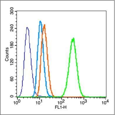
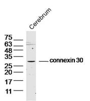
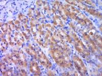
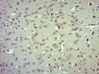
| 产品图片 | 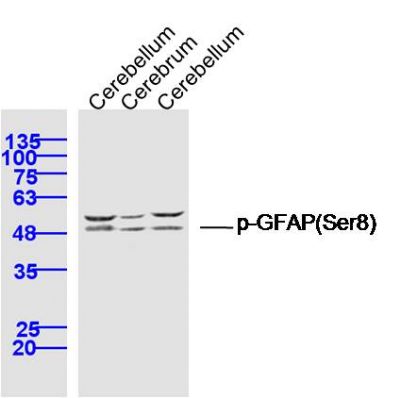 Sample: Cerebellum (Rat) Lysate at 40 ug Cerebrum (Mouse) Lysate at 40 ug Cerebellum (Mouse) Lysate at 40 ug Primary: Anti- phospho-GFAP (Ser8) (bs-5355R) at 1/300 dilution Secondary: IRDye800CW Goat Anti-Rabbit IgG at 1/20000 dilution Predicted band size: 48 kD Observed band size: 48 kD  Paraformaldehyde-fixed, paraffin embedded (Mouse brain); Antigen retrieval by boiling in sodium citrate buffer (pH6.0) for 15min; Block endogenous peroxidase by 3% hydrogen peroxide for 20 minutes; Blocking buffer (normal goat serum) at 37°C for 30min; Antibody incubation with (p-GFAP (Ser8)) Polyclonal Antibody, Unconjugated (bs-5355R) at 1:400 overnight at 4°C, followed by operating according to SP Kit(Rabbit) (sp-0023) instructionsand DAB staining.  Blank control (blue line): hela (fixed with 80% methanol (5 min at -20℃) and then permeabilized with 0.1% PBS-Tween for 20 min at room temperature ). Primary Antibody (green line): Rabbit Anti-GFAP antibody (bs-5355R),Dilution: 3μg /10^6 cells; Isotype Control Antibody (orange line): Rabbit IgG . Secondary Antibody (white blue line): Goat anti-rabbit IgG-FITC,Dilution: 1μg /test. |
风险提示:丁香通仅作为第三方平台,为商家信息发布提供平台空间。用户咨询产品时请注意保护个人信息及财产安全,合理判断,谨慎选购商品,商家和用户对交易行为负责。对于医疗器械类产品,请先查证核实企业经营资质和医疗器械产品注册证情况。
 文献和实验
文献和实验Optimized Protocol to Make Phospho-Specific Antibodies that Work
, not simply its level of expression. In this review, we will discuss both the design of the phosphopeptide immunogen and immunization. The affinity purification of the phospho-specific antibody as well as the methods most suitable for characterizing
Using Phospho‐Motif Antibodies to Determine Kinase Substrates
comprising both the phosphorylated residue and the surrounding residues that determine kinase specificity, with degenerate residues taking up the remaining positions. Currently, several categories of phospho?motif antibody are commercially available
Absorption Control in Immunohistochemistry Using Phospho-Peptides Immobilized on Magnetic Beads
neutralization of phospho-specific antibodies with phospho-peptides immobilized on magnetic beads. This technique allows for sequestration of antibody–peptide complex from the incubation solution, minimizing the risk of formation of unblocked antibodies capable
 技术资料
技术资料暂无技术资料 索取技术资料

