

- ¥1380 - 2200
- LMAI Bio
- LM-11268R
- 进口/国产
- 2025年07月14日
- ELISA=1:500-1000 IHC-P=1:400-800 IHC-F=1:400-800 ICC=1:100-500 IF=1:100-500 (石蜡切片需做抗原修复)
- Rabbit
- Human, Mouse, Rat, Dog, Cow, Horse, Rabbit, Sheep,

企业认证
相关产品推荐更多 >


万千商家帮你免费找货
0 人在求购买到急需产品
- 详细信息
- 文献和实验
- 技术资料
- 供应商:
上海联迈生物工程有限公司
- 库存:
大量
- 目录编号:
LM-11268R
- 克隆性:
多克隆
- 抗原来源:
Rabbit
- 保质期:
1年
- 抗体英文名:
DERLIN-1
- 抗体名:
细胞膜蛋白Derlin抗体
- 宿主:
Rabbit
- 适应物种:
Human, Mouse, Rat, Dog, Cow, Horse, Rabbit, Sheep,
- 免疫原:
KLH conjugated synthetic peptide derived from human DERLIN-1:111-220/251
- 亚型:
IgG
- 形态:
Lyophilized or Liquid
- 应用范围:
ELISA=1:500-1000 IHC-P=1:400-800 IHC-F=1:400-800 ICC=1:100-500 IF=1:100-500 (石蜡切片需做抗原修复)
- 浓度:
1mg/ml
- 保存条件:
Store at -20 °C
- 规格:
100ul 200ul
| 英文名称 | DERLIN-1 |
| 中文名称 | 细胞膜蛋白Derlin抗体 |
| 别 名 | Derlin1; Derlin1; Derlin 1; DER1; DER-1; DER 1; DERL1_HUMAN. |
| 规格价格 | 100ul/1380元 购买 200ul/2200元 购买 大包装/询价 |
| 说 明 书 | 100ul 200ul |
| 研究领域 | 细胞生物 细胞膜蛋白 |
| 抗体来源 | Rabbit |
| 克隆类型 | Polyclonal |
| 交叉反应 | Human, Mouse, Rat, Dog, Cow, Horse, Rabbit, Sheep, |
| 产品应用 | ELISA=1:500-1000 IHC-P=1:400-800 IHC-F=1:400-800 ICC=1:100-500 IF=1:100-500 (石蜡切片需做抗原修复) not yet tested in other applications. optimal dilutions/concentrations should be determined by the end user. |
| 分 子 量 | 29kDa |
| 细胞定位 | 细胞浆 细胞膜 |
| 性 状 | Lyophilized or Liquid |
| 浓 度 | 1mg/ml |
| 免 疫 原 | KLH conjugated synthetic peptide derived from human DERLIN-1:111-220/251 |
| 亚 型 | IgG |
| 纯化方法 | affinity purified by Protein A |
| 储 存 液 | 0.01M TBS(pH7.4) with 1% BSA, 0.03% Proclin300 and 50% Glycerol. |
| 保存条件 | Store at -20 °C for one year. Avoid repeated freeze/thaw cycles. The lyophilized antibody is stable at room temperature for at least one month and for greater than a year when kept at -20°C. When reconstituted in sterile pH 7.4 0.01M PBS or diluent of antibody the antibody is stable for at least two weeks at 2-4 °C. |
| PubMed | PubMed |
| 产品介绍 | background: Degradation in endoplasmic reticulum proteins, also designated Derlins or DERtrins, are crucial for the degradation of misfolded endoplasmic reticulum (ER) luminal proteins. Derlin proteins are multi-pass membrane proteins localizing to the ER. Derlins are involved in transferring misfolded proteins from the ER to the cytosol, where the misfolded proteins are destroyed in an ubiquitin-dependent manner by the proteasome. In the case of cytomegalovirus infection, Derlin-1, as opposed to Derlins-2 and -3, is involved in the export of MHC class I heavy chains from the ER via its interaction with the viral protein US11. Derlins may also be important for cell proliferation. Function: Functional component of endoplasmic reticulum-associated degradation (ERAD) for misfolded lumenal proteins. May act by forming a channel that allows the retrotranslocation of misfolded proteins into the cytosol where they are ubiquitinated and degraded by the proteasome. May mediate the interaction between VCP and the degradation substrate. In case of infection by cytomegaloviruses, it plays a central role in the export from the ER and subsequent degradation of MHC class I heavy chains via its interaction with US11 viral protein, which recognizes and associates with MHC class I heavy chains. Also participates in the degradation process of misfolded cytomegalovirus US2 protein. Subunit: Forms homo- and heterooligomers with DERL2 and DERL3; binding to DERL3 is poorer than that between DERL2 and DERL3. Interacts with AMFR, VIMP/SELS, SEL1L, SYVN1 and VCP, as well as with SEL1L-SYVN1 and VCP-VIMP protein complexes; this interaction is weaker than that observed between DERL2 and these complexes. Interacts with the cytomegalovirus US11 protein. Interacts with NGLY1 and YOD1. Does not bind to EDEM1. Interacts with RNF103. Subcellular Location: Endoplasmic reticulum membrane; Multi-pass membrane protein. Tissue Specificity: Ubiquitous. Similarity: Belongs to the derlin family. SWISS: Q9BUN8 Gene ID: 79139 Database links: Entrez Gene: 420350 Chicken Entrez Gene: 404121 Cow Entrez Gene: 475086 Dog Entrez Gene: 79139 Human Entrez Gene: 67819 Mouse Entrez Gene: 100626802 Pig Entrez Gene: 362912 Rat Omim: 608813 Human SwissProt: Q71SS4 Cow SwissProt: Q9BUN8 Human SwissProt: Q99J56 Mouse Unigene: 241576 Human Unigene: 289387 Mouse Unigene: 110990 Rat Important Note: This product as supplied is intended for research use only, not for use in human, therapeutic or diagnostic applications. |
| 产品图片 |
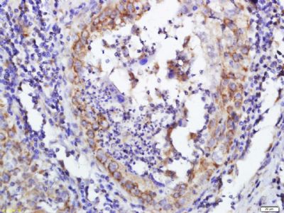
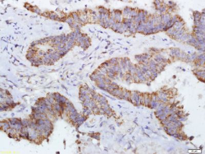
Tissue/cell: human esophageal carcinoma; 4% Paraformaldehyde-fixed and paraffin-embedded;
Antigen retrieval: citrate buffer ( 0.01M, pH 6.0 ), Boiling bathing for 15min; Block endogenous peroxidase by 3% Hydrogen peroxide for 30min; Blocking buffer (normal goat serum,C-0005) at 37℃ for 20 min; Incubation: Anti-DERLIN-1 Polyclonal Antibody, Unconjugated(bs-11268R) 1:600, overnight at 4°C, followed by conjugation to the secondary antibody(SP-0023) and DAB(C-0010) staining 
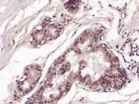
Tissue/cell: human colon carcinoma; 4% Paraformaldehyde-fixed and paraffin-embedded;
Antigen retrieval: citrate buffer ( 0.01M, pH 6.0 ), Boiling bathing for 15min; Block endogenous peroxidase by 3% Hydrogen peroxide for 30min; Blocking buffer (normal goat serum,C-0005) at 37℃ for 20 min; Incubation: Anti-DERLIN-1 Polyclonal Antibody, Unconjugated(bs-11268R) 1:600, overnight at 4°C, followed by conjugation to the secondary antibody(SP-0023) and DAB(C-0010) staining |
风险提示:丁香通仅作为第三方平台,为商家信息发布提供平台空间。用户咨询产品时请注意保护个人信息及财产安全,合理判断,谨慎选购商品,商家和用户对交易行为负责。对于医疗器械类产品,请先查证核实企业经营资质和医疗器械产品注册证情况。
 文献和实验
文献和实验[2] ✦ CUT&RUN 原理: 图 4:CUT&RUN 原理 [3] 利用连有刀豆蛋白 A 的磁珠(concanavalin A-coated magnetic beads)结合细胞。使用非离子去污剂洋地黄皂苷进行细胞膜通透。然后孵育靶蛋白(如转录因子, TF )的抗体和 Protein A-MNase 。抗体和 Protein A-MNase 能够通过核孔进入细胞核,MNase 通过 Protein A 和抗体的介导切割靶蛋白附近的 DNA 序列。 MNase 的活化需要 Ca
在胞质中以单体形式存在,发出绿色的荧光。可以在荧光显微镜下,或流式检测。采用488nm激发,其检测波长分别是527nm和590nm。整个实验过程操作简单,只需要30min就可以见到结果。Caspase家族可以检测的分子非常多,也有不少商业的试剂盒可以应用。即使没有相应的试剂盒,只要有相应抗体基本上是可以检测的,具体的方法是参照细胞内蛋白检测的步骤。在细胞凋亡过程中伴随着一系列的形态特征改变,细胞膜的改变是这些特征中较早出现的一种。在凋亡细胞中,细胞膜磷脂酰丝氨酸(PS)从细胞膜的内侧翻转到细胞膜
血型系统。此外,还有一些抗原,或因其在群体中出现的频率太高,或因其在群体中分布的频率太低,对它们无法进行遗传学分析。在没有弄清它们的遗传关系以前,暂且把这些抗原分别叫做高频率抗原及低频率抗原,对于它们的归属有待进一步确定。 红细胞血型抗原 红细胞膜中夹杂着3种蛋白质:糖蛋白、简单蛋白及膜收缩蛋白。红细胞抗原有些突出在细胞表面,好像伸出在地面上的树枝,如ABH抗原;有些镶嵌在细胞膜内,如Rh抗原。抗原与抗体发生特异反应的部分,叫做抗原决定簇。血型抗原决定簇的化学组成,有的已经清楚,但大部分不清楚。有些血型
 技术资料
技术资料暂无技术资料 索取技术资料

