


- ¥1380 - 2200
- 康朗生物
- kl-20593R
- 中国/美国/德国
- 2025年07月15日
- ELISA=1:500-1000 IHC-P=1:400-800 IHC-F=1:400-800 Flow-Cyt=1μg/Test ICC=1:100-500 IF=1:100-500
- Rabbit
- Human, Mouse, Rat, Dog, Pig, Rabbit, Sheep,

企业认证
相关产品推荐更多 >

万千商家帮你免费找货
0 人在求购买到急需产品
- 详细信息
- 文献和实验
- 技术资料
- 供应商:
上海康朗生物科技有限公司
- 库存:
大量
- 目录编号:
kl-20593R
- 克隆性:
多克隆
- 抗原来源:
Rabbit
- 保质期:
12个月
- 抗体英文名:
PKC delta antibody
- 抗体名:
蛋白激酶C亚性D型抗体
- 宿主:
Rabbit
- 适应物种:
Human, Mouse, Rat, Dog, Pig, Rabbit, Sheep,
- 免疫原:
KLH conjugated synthetic peptide derived from human PKC delta:1-100/676
- 亚型:
IgG
- 形态:
冻干粉或液体
- 应用范围:
ELISA=1:500-1000 IHC-P=1:400-800 IHC-F=1:400-800 Flow-Cyt=1μg/Test ICC=1:100-500 IF=1:100-500
- 浓度:
1mg/ml
- 保存条件:
-20 °C
- 规格:
100ul 200ul
| 中文名称 | 蛋白激酶C亚性D型抗体 |
| 别 名 | MAY 1; MAY1; nPKC delta; PCKd; PKC d; PKC delta; PKC-d; PKCD; PKCdelta; PRKC D; PRKC delta; PRKC-d; PRKCd; Protein Kinase C delta; Protein kinase C delta type; Protein Kinase Cdelta; KPCD_HUMAN. |
| 规格价格 | 100ul/1380元 购买 200ul/2200元 购买 大包装/询价 |
| 说 明 书 | 100ul 200ul |
| 研究领域 | 肿瘤 细胞生物 免疫学 细胞凋亡 转录调节因子 激酶和磷酸酶 |
| 抗体来源 | Rabbit |
| 克隆类型 | Polyclonal |
| 交叉反应 | Human, Mouse, Rat, Dog, Pig, Rabbit, Sheep, |
| 产品应用 | ELISA=1:500-1000 IHC-P=1:400-800 IHC-F=1:400-800 Flow-Cyt=1μg/Test ICC=1:100-500 IF=1:100-500 (石蜡切片需做抗原修复) not yet tested in other applications. optimal dilutions/concentrations should be determined by the end user. |
| 分 子 量 | 77kDa |
| 细胞定位 | 细胞核 细胞浆 细胞膜 |
| 性 状 | Lyophilized or Liquid |
| 浓 度 | 1mg/ml |
| 免 疫 原 | KLH conjugated synthetic peptide derived from human PKC delta:1-100/676 |
| 亚 型 | IgG |
| 纯化方法 | affinity purified by Protein A |
| 储 存 液 | 0.01M TBS(pH7.4) with 1% BSA, 0.03% Proclin300 and 50% Glycerol. |
| 保存条件 | Store at -20 °C for one year. Avoid repeated freeze/thaw cycles. The lyophilized antibody is stable at room temperature for at least one month and for greater than a year when kept at -20°C. When reconstituted in sterile pH 7.4 0.01M PBS or diluent of antibody the antibody is stable for at least two weeks at 2-4 °C. |
| PubMed | PubMed |
| 产品介绍 | background: Protein kinase C (PKC) is a family of serine- and threonine-specific protein kinases that can be activated by calcium and the second messenger diacylglycerol. PKC family members phosphorylate a wide variety of protein targets and are known to be involved in diverse cellular signaling pathways. PKC family members also serve as major receptors for phorbol esters, a class of tumor promoters. Each member of the PKC family has a specific expression profile and is believed to play distinct roles in cells. The protein encoded by this gene is one of the PKC family members. Studies both in human and mice demonstrate that this kinase is involved in B cell signaling and in the regulation of growth, apoptosis, and differentiation of a variety of cell types. Alternatively spliced transcript variants encoding the same protein have been observed. [provided by RefSeq, Jul 2008]. Function: Calcium-independent, phospholipid- and diacylglycerol (DAG)-dependent serine/threonine-protein kinase that plays contrasting roles in cell death and cell survival by functioning as a pro-apoptotic protein during DNA damage-induced apoptosis, but acting as an anti-apoptotic protein during cytokine receptor-initiated cell death, is involved in tumor suppression as well as survival of several cancers, is required for oxygen radical production by NADPH oxidase and acts as positive or negative regulator in platelet functional responses. Upon DNA damage, activates the promoter of the death-promoting transcription factor BCLAF1/Btf to trigger BCLAF1-mediated p53/TP53 gene transcription and apoptosis. In response to oxidative stress, interact with and activate CHUK/IKKA in the nucleus, causing the phosphorylation of p53/TP53. In the case of ER stress or DNA damage-induced apoptosis, can form a complex with the tyrosine-protein kinase ABL1 which trigger apoptosis independently of p53/TP53. In cytosol can trigger apoptosis by activating MAPK11 or MAPK14, inhibiting AKT1 and decreasing the level of X-linked inhibitor of apoptosis protein (XIAP), whereas in nucleus induces apoptosis via the activation of MAPK8 or MAPK9. Upon ionizing radiation treatment, is required for the activation of the apoptosis regulators BAX and BAK, which trigger the mitochondrial cell death pathway. Can phosphorylate MCL1 and target it for degradation which is sufficient to trigger for BAX activation and apoptosis. Is required for the control of cell cycle progression both at G1/S and G2/M phases. Mediates phorbol 12-myristate 13-acetate (PMA)-induced inhibition of cell cycle progression at G1/S phase by up-regulating the CDK inhibitor CDKN1A/p21 and inhibiting the cyclin CCNA2 promoter activity. In response to UV irradiation can phosphorylate CDK1, which is important for the G2/M DNA damage checkpoint activation. Can protect glioma cells from the apoptosis induced by TNFSF10/TRAIL, probably by inducing increased phosphorylation and subsequent activation of AKT1. Is highly expressed in a number of cancer cells and promotes cell survival and resistance against chemotherapeutic drugs by inducing cyclin D1 (CCND1) and hyperphosphorylation of RB1, and via several pro-survival pathways, including NF-kappa-B, AKT1 and MAPK1/3 (ERK1/2). Can also act as tumor suppressor upon mitogenic stimulation with PMA or TPA. In N-formyl-methionyl-leucyl-phenylalanine (fMLP)-treated cells, is required for NCF1 (p47-phox) phosphorylation and activation of NADPH oxidase activity, and regulates TNF-elicited superoxide anion production in neutrophils, by direct phosphorylation and activation of NCF1 or indirectly through MAPK1/3 (ERK1/2) signaling pathways. May also play a role in the regulation of NADPH oxidase activity in eosinophil after stimulation with IL5, leukotriene B4 or PMA. In collagen-induced platelet aggregation, acts a negative regulator of filopodia formation and actin polymerization by interacting with and negatively regulating VASP phosphorylation. Downstream of PAR1, PAR4 and CD36/GP4 receptors, regulates differentially platelet dense granule secretion; acts as a positive regulator in PAR-mediated granule secretion, whereas it negatively regulates CD36/GP4-mediated granule release. Phosphorylates MUC1 in the C-terminal and regulates the interaction between MUC1 and beta-catenin. Subunit: Interacts with PDK1 (via N-terminus region), RAD9A, CDCP1, MUC1 and VASP. Subcellular Location: Cytoplasm. Cytoplasm, perinuclear region. Nucleus. Endoplasmic reticulum. Mitochondrion. Cell membrane; Peripheral membrane protein. Post-translational modifications: Autophosphorylated and/or phosphorylated at Thr-507, within the activation loop; phosphorylation at Thr-507 is not a prerequisite for enzymatic activity. Autophosphorylated at Ser-299, Ser-302 and Ser-304. Upon TNFSF10/TRAIL treatment, phosphorylated at Tyr-155; phosphorylation is required for its translocation to the endoplasmic reticulum and cleavage by caspase-3. Phosphorylated at Tyr-313, Tyr-334 and Tyr-567; phosphorylation of Tyr-313 and Tyr-567 following thrombin stimulation potentiates its kinase activity. Phosphorylated by protein kinase PDK1; phosphorylation is inhibited by the apoptotic C-terminus cleavage product of PKN2. Similarity: Belongs to the protein kinase superfamily. AGC Ser/Thr protein kinase family. PKC subfamily. Contains 1 AGC-kinase C-terminal domain. Contains 1 C2 domain. Contains 2 phorbol-ester/DAG-type zinc fingers. Contains 1 protein kinase domain. SWISS: Q05655 Gene ID: 5580 Database links: Entrez Gene: 5580 Human Entrez Gene: 18753 Mouse Entrez Gene: 170538 Rat Omim: 176977 Human SwissProt: Q05655 Human SwissProt: P28867 Mouse SwissProt: P09215 Rat Unigene: 155342 Human Unigene: 2314 Mouse Unigene: 98279 Rat Important Note: This product as supplied is intended for research use only, not for use in human, therapeutic or diagnostic applications. |
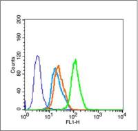
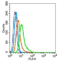
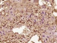
| 产品图片 | 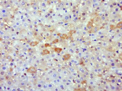 Paraformaldehyde-fixed, paraffin embedded (Rat liver); Antigen retrieval by boiling in sodium citrate buffer (pH6.0) for 15min; Block endogenous peroxidase by 3% hydrogen peroxide for 20 minutes; Blocking buffer (normal goat serum) at 37°C for 30min; Antibody incubation with (PKC delta) Polyclonal Antibody, Unconjugated (bs-20593R) at 1:500 overnight at 4°C, followed by a conjugated secondary (sp-0023) for 20 minutes and DAB staining. 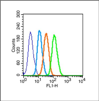 Blank control (Black line): Raji (Black). Primary Antibody (green line): Rabbit Anti-PKC delta (Ser32) antibody (bs-20593R) Dilution: 1μg /10^6 cells; Isotype Control Antibody (orange line): Rabbit IgG . Secondary Antibody (white blue line): Goat anti-rabbit IgG-PE Dilution: 1μg /test. Protocol The cells were fixed with 4% PFA (10min)and then permeabilized with 90% ice-cold methanol for 20 min on ice. Cells stained with Primary Antibody for 30 min at room temperature. The cells were then incubated in 1 X PBS/2%BSA/10% goat serum to block non-specific protein-protein interactions followed by the antibody for 15 min at room temperature. The secondary antibody used for 40 min at room temperature. Acquisition of 20,000 events was performed. |
风险提示:丁香通仅作为第三方平台,为商家信息发布提供平台空间。用户咨询产品时请注意保护个人信息及财产安全,合理判断,谨慎选购商品,商家和用户对交易行为负责。对于医疗器械类产品,请先查证核实企业经营资质和医疗器械产品注册证情况。
 文献和实验
文献和实验licanming 我的实验是用VEGF处理细胞后测定细胞PKC激活水平,拟采用Western blot的方法,用ECL法检测,这样分析PKC激活程度结果国际上认可吗? 我在Cell Signaling Technology上,查到 2261 Phospho-(Ser) PKC Substrate Antibody W IP E D 9371 Phospho-PKC (pan) (betaII Ser660) Antibody W IP
上海西唐生物科技有限公司 021-55229872, 65333639 www.westang.com 大鼠蛋白激酶 C(PKC)ELISA 试剂盒 ( 用于血清、血浆、细胞培养上清液和其它生物体液内 ) 原理 本实验采用双抗体夹心 ABC-ELISA 法。用抗大鼠 PKC 单抗包被于酶标板上,标准品和样品中的 PKC 与单抗结合,加入生物素化的抗大鼠 PKC ,形成免疫复合物连接在板上,辣根
上海西唐生物科技有限公司 021-55229872, 65333639 www.westang.com 人蛋白激酶C (PKC)ELISA 试剂盒 ( 用于血清、血浆、细胞培养上清液和其它生物体液内 ) 原理 本实验采用双抗体夹心 ABC-ELISA 法。用抗人 PKC 单抗包被于酶标板上,标准品和样品中的 PKC与单抗结合,加入生物素化的抗人 PKC ,形成免疫复合物连接
 技术资料
技术资料暂无技术资料 索取技术资料
