

- ¥1380 - 2200
- LMAI Bio
- LM-0018R
- 进口/国产
- 2026年05月27日
- WB=1:500-2000 ELISA=1:500-1000 IHC-P=1:400-800 IHC-F=1:400-800 IF=1:100-500 (石蜡切片需做抗原修复)
- Rabbit
- Human, Mouse, Rat, Chicken, Pig, Cow,

企业认证
相关产品推荐更多 >


万千商家帮你免费找货
0 人在求购买到急需产品
- 详细信息
- 文献和实验
- 技术资料
- 供应商:
上海联迈生物工程有限公司
- 库存:
大量
- 目录编号:
LM-0018R
- 克隆性:
多克隆
- 抗原来源:
Rabbit
- 保质期:
1年
- 抗体英文名:
IDE
- 抗体名:
胰岛素降解酶抗体
- 宿主:
Rabbit
- 适应物种:
Human, Mouse, Rat, Chicken, Pig, Cow,
- 免疫原:
KLH conjugated synthetic peptide derived from human IDE:491-590/1019
- 亚型:
IgG
- 形态:
Lyophilized or Liquid
- 应用范围:
WB=1:500-2000 ELISA=1:500-1000 IHC-P=1:400-800 IHC-F=1:400-800 IF=1:100-500 (石蜡切片需做抗原修复)
- 浓度:
1mg/ml
- 保存条件:
Store at -20 °C
- 规格:
100ul 200ul
| 中文名称 | 胰岛素降解酶抗体 |
| 别 名 | BC2; Insulin degrading enzyme; FLJ35968; insulin protease; insulinase; insulysin; Abeta-degrading protease; FLJ35968; Ide; IDE_HUMAN; Insulin-degrading enzyme; OTTHUMP00000020097. |
| 规格价格 | 100ul/1380元 购买 200ul/2200元 购买 大包装/询价 |
| 说 明 书 | 100ul 200ul |
| 研究领域 | 心血管 细胞生物 免疫学 神经生物学 信号转导 生长因子和激素 合成与降解 糖尿病 |
| 抗体来源 | Rabbit |
| 克隆类型 | Polyclonal |
| 交叉反应 | Human, Mouse, Rat, Chicken, Pig, Cow, |
| 产品应用 | WB=1:500-2000 ELISA=1:500-1000 IHC-P=1:400-800 IHC-F=1:400-800 IF=1:100-500 (石蜡切片需做抗原修复) not yet tested in other applications. optimal dilutions/concentrations should be determined by the end user. |
| 分 子 量 | 54/117kDa |
| 细胞定位 | 细胞浆 细胞膜 分泌型蛋白 |
| 性 状 | Lyophilized or Liquid |
| 浓 度 | 1mg/ml |
| 免 疫 原 | KLH conjugated synthetic peptide derived from human IDE:491-590/1019 |
| 亚 型 | IgG |
| 纯化方法 | affinity purified by Protein A |
| 储 存 液 | 0.01M TBS(pH7.4) with 1% BSA, 0.03% Proclin300 and 50% Glycerol. |
| 保存条件 | Store at -20 °C for one year. Avoid repeated freeze/thaw cycles. The lyophilized antibody is stable at room temperature for at least one month and for greater than a year when kept at -20°C. When reconstituted in sterile pH 7.4 0.01M PBS or diluent of antibody the antibody is stable for at least two weeks at 2-4 °C. |
| PubMed | PubMed |
| 产品介绍 | background: Insulysin was identified nearly a century ago as an enzyme responsible for the degradation of insulin in cells, although the precise interactions between insulin and insulysin remain elusive. Human insulysin was cloned in 1988, and shown to be a 118 kDa protein that exists primarily as a homodimer, and perhaps also complexed with other molecules. The sequence is well conserved between humans, rats and mice, and the antibody recognizes these species. Insulysin is a metalloproteinase of the clan ME, family M16, which contains an active site HxxEH, a reversal of the canonical HExxH zinc binding motif. Considered a zinc metalloproteinase, the activity of insulysin can be blocked with EDTA or 1-10 phenanthroline. In addition to the active metalloproteinase domain, insulysin contains a second metalloproteinase site which is considered catalytically inactive, and is thought to assist in substrate binding. Insulysin is most closely related to the bacterial proteinase pitrilysin, (the human orthologue of which appears to be MPRP1) and the mammalian proteinsae nardilysin. Generally thought to be a cytoplasmic protein, insulysin has been isolated from many different tissues and cell lines, and can degrade intact insulin, insulin B chain, glucagon, denatured hemoglobin, alpha amyloid protein, TGF alpha and amylin. Recent work implicates insulysin in clearing beta amyloid plaques from the brain, and has generated much interest in Alzheimer’s disease research. The pH optimum for insulysin is basic, pH 8.5, which also distinguishes it from other metalloproteinases. Function: Plays a role in the cellular breakdown of insulin, IAPP, glucagon, bradykinin, kallidin and other peptides, and thereby plays a role in intercellular peptide signaling. Degrades amyloid formed by APP and IAPP. May play a role in the degradation and clearance of naturally secreted amyloid beta-protein by neurons and microglia. Subunit: Homodimer. Can form higher oligomers. Interacts (via N-terminus) with varicella-zoster virus (VZV) envelope glycoprotein E (via N-terminus); the membrane-associated isoform may function as an entry receptor for this virus. Subcellular Location: Cytoplasm. Cell membrane. Secreted. Note=Present at the cell surface of neuron cells. The membrane-associated isoform is approximately 5 kDa larger than the known cytosolic isoform. Post-translational modifications: The N-terminus is blocked. Similarity: Belongs to the peptidase M16 family. SWISS: P14735 Gene ID: 3416 Database links: Entrez Gene: 3416 Human Entrez Gene: 15925 Mouse Entrez Gene: 25700 Rat Omim: 146680 Human SwissProt: P14735 Human SwissProt: Q9JHR7 Mouse SwissProt: P35559 Rat Unigene: 500546 Human Unigene: 28366 Mouse Unigene: 45029 Rat Important Note: This product as supplied is intended for research use only, not for use in human, therapeutic or diagnostic applications. |
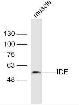
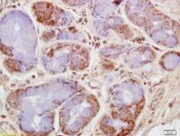
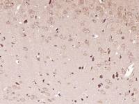
| 产品图片 | 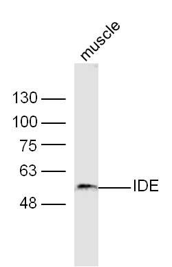 Sample: Muscle (Mouse) Lysate at 40 ug Primary: Anti-IDE (bs-0018R) at 1/300 dilution Secondary: IRDye800CW Goat Anti-Rabbit IgG at 1/20000 dilution Predicted band size: 117 kD Observed band size: 54 kD 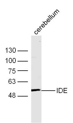 Sample: Cerebellum (Mouse) Lysate at 40 ug Primary: Anti-IDE (bs-0018R) at 1/300 dilution Secondary: IRDye800CW Goat Anti-Rabbit IgG at 1/20000 dilution Predicted band size: 117 kD Observed band size: 54 kD 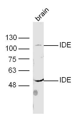 Sample: Brain (Mouse) Lysate at 40 ug Primary: Anti-IDE (bs-0018R) at 1/300 dilution Secondary: IRDye800CW Goat Anti-Rabbit IgG at 1/20000 dilution Predicted band size: 117 kD Observed band size: 54/117 kD 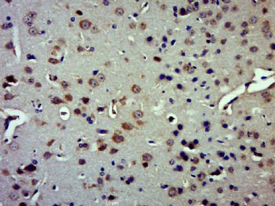 Paraformaldehyde-fixed, paraffin embedded (Mouse brain); Antigen retrieval by boiling in sodium citrate buffer (pH6.0) for 15min; Block endogenous peroxidase by 3% hydrogen peroxide for 20 minutes; Blocking buffer (normal goat serum) at 37°C for 30min; Antibody incubation with (IDE) Polyclonal Antibody, Unconjugated (bs-0018R) at 1:500 overnight at 4°C, followed by a conjugated secondary (sp-0023) for 20 minutes and DAB staining. |
风险提示:丁香通仅作为第三方平台,为商家信息发布提供平台空间。用户咨询产品时请注意保护个人信息及财产安全,合理判断,谨慎选购商品,商家和用户对交易行为负责。对于医疗器械类产品,请先查证核实企业经营资质和医疗器械产品注册证情况。
 文献和实验
文献和实验练的是肌肉,为啥全身都受益?新研究揭示运动激活全身代谢的关键
的一种蛋白在肝脏中激活重要的细胞「自噬」过程,使肝脏的代谢功能更强。 根据论文,研究人员在实验小鼠运动前后对它们的血液进行了蛋白质组学分析,由此发现,运动诱导骨骼肌分泌一种叫做纤维粘连蛋白(FN1)的可溶性蛋白质。 而这些 FN1 蛋白随着血液循环到达肝脏后,通过肝细胞表面的一种受体整合素 α5β1 发出信号,在肝脏中激活「细胞自噬」过程,促进代谢适应。细胞自噬,简单来说就是细胞内的一种物质回收机制,降解和清除细胞内多余的蛋白质或受损的细胞器等。因此,运动在肝脏中激活细胞自噬也意味着这个代谢器官
Cell Metabo: 另辟蹊径!武汉大学李红良等团队合作发现 2 型糖尿病治疗新靶点
2018 年 the Lancet 研究统计显示,空腹高血糖是中国人群寿命的第三大危险因素 (1)。人体 90% 的内源性葡萄糖来自肝葡萄糖生成(hepatic glucose production, HGP),是维持体内糖稳态的重要组成部分 (2)。 图片来源:the Lancet 空腹 HGP 增加是 T2D (2 型糖尿病)的标志,HGP 率高于正常生理水平,表现为肝胰岛素抵抗,葡萄糖代谢能力下降,是患者空腹高血糖的一个重要原因。现有的降糖药物对 T2D 患者有疗效,但仍有局限性。令人
分泌,另一方面抑制胰高血糖素释放,既能降低餐后血糖,还能延缓胃排空、增强饱腹感。但DPP4会快速降解这些宝贵的肠促胰岛素激素,让其血糖调节作用大打折扣。 02 DPP4 抑制剂的核心作用 糖尿病被称为“牵一发而动全身”的疾病,其危害不仅限于血糖控制不佳,而是对全身多个器官和系统造成渐进性的、难以察觉的影响,其中2型糖尿病(T2DM)占糖尿病比例超90%,患者多面临胰岛B细胞功能渐进下降、胰岛素分泌不足或胰岛素抵抗的问题。 DPP4抑制剂的出现,为血糖控制提供了新思路——这类新型口服降糖药(如西格
 技术资料
技术资料暂无技术资料 索取技术资料

