


- ¥2800
- wksubio
- 不限
- 国内
- SU-F00021
- 2026年05月29日

企业认证
相关产品推荐更多 >


万千商家帮你免费找货
0 人在求购买到急需产品
- 详细信息
- 文献和实验
- 技术资料
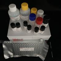
- 库存:
大量
- 供应商:
上海瓦兰生物
- 检测范围:
不限
- 检测方法:
酶联免疫法
- 应用:
科研单位
- 适应物种:
不限
- 标记物:
呋喃它酮
- 样本:
液体
- 规格:
50t
| Name | 96 determinations | 48 determinations |
| Microelisa stripplate | 12*8strips | 12*4strips |
| Standard | 0.3ml | 0.3ml |
| Sample diluent | 6.0ml | 3.0ml |
| HRP-Conjugate reagent | 10.0ml | 5.0ml |
| 20X Wash solution | 25ml | 15ml |
| Chromogen Solution A | 6.0ml | 3.0ml |
| Chromogen Solution B | 6.0ml | 3.0ml |
| Stop Solution | 6.0ml | 3.0ml |
| Closure plate membrane | 2 | 2 |
| User manual | 1 | 1 |
| Sealed bags | 1 | 1 |
8、4、2、1、0.5、0 ng/ml.
Reagent preparation
20×wash solution:Dilute with Distilled or deionized water 1:20.
Assay procedure
1. Prepare all reagents before starting assay procedure. It is recommended that all Standards and Samples be added in duplicate to the Microelisa Stripplate.
2. Add standard: Set Standard wells, testing sample wells. Add standard 50μl to standard well.
3. Add Sample: Add testing sample 10μl Then add sample diluent 40μl to testing sample well; Blank well doesn’t add anyting.
4. Add 100μl of HRP-conjugate reagent to each well, cover with an adhesive strip and incubate for 60 minutes at 37°C.
5. Aspirate each well and wash, repeating the process four times for a total of five washes. Wash by filling each well with Wash Solution (400μl) using a squirt bottle, manifold dispenser or autowasher. Complete removal of liquid at each step is essential to good performance. After the last wash, remove any remaining Wash Solution by aspirating or decanting. Invert the plate and blot it against clean paper towels.
6. Add chromogen solution A 50μl and chromogen solution B 50μl to each well. Gently mix and incubate for 15 minutes at 37°C. Protect from light.
7. Add 50μl Stop Solution to each well. The color in the wells should change from blue to yellow. If the color in the wells is green or the color change does not
appear uniform, gently tap the plate to ensure thorough mixing.
8. Read the Optical Density (O.D.) at 450 nm using a microtiter plate reader within 15 minutes.
Calculation of results
- This standard curve is used to determine the amount in an unknown sample. The standard curve is generated by plotting the average O.D. (450 nm) obtained for each of the six standard concentrations on the vertical (Y) axis versus the corresponding concentration on the horizontal (X) axis.
- First, calculate the mean O.D. value for each standard and sample. All O.D. values, are subtracted by the mean value of the zero standard before result interpretation. Construct the standard curve using graph paper or statistical software.
- To determine the amount in each sample, first locate the O.D. value on the Y-axis and extend a horizontal line to the standard curve. At the point of intersection, draw a vertical line to the X-axis and read the corresponding concentration.
- Any variation in operator, pipetting and washing technique, incubation time or temperature, and kit age can cause variation in result. Each user should obtain their own standard curve.
- The sensitivity by this assay is 0.1 ng/ml.
- Standard curve
Storage: 2-8℃.
validity: six months.
FOR RESEARCH USE ONLY; NOT FOR THERAPEUTIC OR DIAGNOSTIC APPLICATIONS! PLEASE READ THROUGH ENTIRE PROCEDURE BEFORE BEGINNING!
兽药残留系列
Serum - Use a serum separator tube and allow samples to clot for 30 minutes before centrifugation for 10 minutes at approximately 3000×g. Remove serum and assay immediately or aliquot and store samples at -20℃ or -80℃.Avoid repeated freeze-thaw cycles
Plasma - Collect plasma using EDTA or heparin as an anticoagulant. Centrifuge samples for 30 minutes at 3000×g at 2-8℃ within 30 minutes of collection. Store samples at -20℃or -80℃. Avoid repeated freeze-thaw cycles.
Cell culture supernates and other biological fluids - Remove particulates by centrifugation and assay immediately or aliquot and store samples at -20℃or -80℃. Avoid repeated freeze-thaw cycles.
Note: The samples should be centrifugated adequately and no hemolysis or granule was allowed.
Materials required but not supplied
1. Standard microplate reader(450nm)
2. Precision pipettes and Disposable pipette tips.
3. 37 ℃ incubator
Precautions
1. Do not substitute reagents from one kit to another. Standard, conjugate and microplates are matched for optimal performance. Use only the reagents supplied by manufacturer.
2. Do not remove microplate from the storage bag until needed. Unused strips should be stored at 2-8°C in their pouch with the desiccant provided.
3. Mix all reagents before using.
Remove all kit reagents from refrigerator and allow them to reach room temperature ( 20-25°C)
Materials supplied
| Name | 96 determinations | 48 determinations |
| Microelisa stripplate | 12*8strips | 12*4strips |
| Standard | 0.3ml | 0.3ml |
| Sample diluent | 6.0ml | 3.0ml |
| HRP-Conjugate reagent | 10.0ml | 5.0ml |
| 20X Wash solution | 25ml | 15ml |
| Chromogen Solution A | 6.0ml | 3.0ml |
| Chromogen Solution B | 6.0ml | 3.0ml |
| Stop Solution | 6.0ml | 3.0ml |
| Closure plate membrane | 2 | 2 |
| User manual | 1 | 1 |
| Sealed bags | 1 | 1 |
8、4、2、1、0.5、0 ng/ml.
Reagent preparation
20×wash solution:Dilute with Distilled or deionized water 1:20.
Assay procedure
1. Prepare all reagents before starting assay procedure. It is recommended that all Standards and Samples be added in duplicate to the Microelisa Stripplate.
2. Add standard: Set Standard wells, testing sample wells. Add standard 50μl to standard well.
3. Add Sample: Add testing sample 10μl Then add sample diluent 40μl to testing sample well; Blank well doesn’t add anyting.
4. Add 100μl of HRP-conjugate reagent to each well, cover with an adhesive strip and incubate for 60 minutes at 37°C.
5. Aspirate each well and wash, repeating the process four times for a total of five washes. Wash by filling each well with Wash Solution (400μl) using a squirt bottle, manifold dispenser or autowasher. Complete removal of liquid at each step is essential to good performance. After the last wash, remove any remaining Wash Solution by aspirating or decanting. Invert the plate and blot it against clean paper towels.
6. Add chromogen solution A 50μl and chromogen solution B 50μl to each well. Gently mix and incubate for 15 minutes at 37°C. Protect from light.
7. Add 50μl Stop Solution to each well. The color in the wells should change from blue to yellow. If the color in the wells is green or the color change does not
appear uniform, gently tap the plate to ensure thorough mixing.
8. Read the Optical Density (O.D.) at 450 nm using a microtiter plate reader within 15 minutes.
Calculation of results
- This standard curve is used to determine the amount in an unknown sample. The standard curve is generated by plotting the average O.D. (450 nm) obtained for each of the six standard concentrations on the vertical (Y) axis versus the corresponding concentration on the horizontal (X) axis.
- First, calculate the mean O.D. value for each standard and sample. All O.D. values, are subtracted by the mean value of the zero standard before result interpretation. Construct the standard curve using graph paper or statistical software.
- To determine the amount in each sample, first locate the O.D. value on the Y-axis and extend a horizontal line to the standard curve. At the point of intersection, draw a vertical line to the X-axis and read the corresponding concentration.
- Any variation in operator, pipetting and washing technique, incubation time or temperature, and kit age can cause variation in result. Each user should obtain their own standard curve.
- The sensitivity by this assay is 0.1 ng/ml.
- Standard curve
Storage: 2-8℃.
validity: six months.
FOR RESEARCH USE ONLY; NOT FOR THERAPEUTIC OR DIAGNOSTIC APPLICATIONS! PLEASE READ THROUGH ENTIRE PROCEDURE BEFORE BEGINNING!
风险提示:丁香通仅作为第三方平台,为商家信息发布提供平台空间。用户咨询产品时请注意保护个人信息及财产安全,合理判断,谨慎选购商品,商家和用户对交易行为负责。对于医疗器械类产品,请先查证核实企业经营资质和医疗器械产品注册证情况。
 文献和实验
文献和实验不显示)。 3.1.4出现BioLIAISON主菜单(在主菜单的VITEK下点击),出现VITEK Status状态框,点击Reader,出现Status和Print,点击Status。 3.1.5 在读数状态窗口击process on钮。开始执行任务。3.2测试标本3.2.1标本的稀释:选取经纯培养18~24小时后,大小为3mm左右的待测菌落2~3个,置于装有1.8ml0.45%生理盐水的试管中进行稀释,用标准比浊计测菌液浓度(如浊度高加生理盐水,浊度低加菌落)。最后的菌液浓度必须达到测试卡
【原理】 肺炎支原体快速检测卡可检测血清中的肺炎支原体lgM抗体。患者的血清样品分别加入两个测试孔内,当样品沿膜扩散时,分别加入3滴抗人lgM碱性磷酸酶复合物、3滴洗液和2滴底物,5分钟后观察结果。质控孔为质量控制,固定有人lgM,测试孔固定有肺炎支原体抗原。阳性反应结果为蓝色,阴性结果为无色。 【材料】 1、患者血清。 2、肺炎支原体lgM检测试剂盒,包括: (1)检测卡:单独锡纸包装,卡上固定有肺炎支原体抗原或人lgM(质控孔)。 (2)阳性质控液(1.7ml
的胶体金检测试产品。如 HCG 测试卡 ,HBV 测试卡等。 Shiach CR 等人用 POCT 在实验室测定凝血酶原时间 ( PT) 发现两组 INR 时间是相似的 ,分别为 60.19 % 和 59.13 % ,调查问卷显示对 POCT 监测病人显示很满意。 四 、选择性电极技术 用离子选择性电极结合传感器包括生物传感器和化学传感器技术,制成了便携式快速检测血气(PH 、 PCO 2 、 PO 2 等)和电解质 (K+ 、 Na+ 、 CL- 等 ) 的仪器,已被广泛应用于临床。 五
 技术资料
技术资料需要更多技术资料 索取更多技术资料

