

- ¥1700 - 4000
- GeneTex

- 美国
- GTX629888
- 2026年02月12日
- WB, ICC/IF
- Mouse
- Human, Rat

企业认证
相关产品推荐更多 >
万千商家帮你免费找货
0 人在求购买到急需产品
- 详细信息
- 文献和实验
- 技术资料
- 免疫原:
Recombinant protein encompassing a sequence within the center region of human SQSTM1 / P62. The exact sequence is proprietary.
- 亚型:
IgG2a
- 形态:
Liquid
- 保存条件:
Store as concentrated solution. Centrifuge briefly prior to opening vial. For short-term storage (1-2 weeks), store at 4ºC. For long-term storage, aliquot and store at -20ºC or below. Avoid multiple freeze-thaw cycles.
- 克隆性:
Monoclonal
- 标记物:
Unconjugated
- 适应物种:
Human, Rat
- 保质期:
12 months from the shipping date of the product.
- 抗原来源:
Human
- 目录编号:
GTX629888
- 级别:
Primary Antibodies
- 库存:
Available
- 供应商:
GeneTex
- 宿主:
Mouse
- 应用范围:
WB, ICC/IF
- 浓度:
1 mg/ml (Please refer to the vial label for the specific concentration.)
- 靶点:
SQSTM1 / P62
- 抗体英文名:
SQSTM1 / P62 antibody [GT239]
- 抗体名:
SQSTM1 / P62 抗体 [GT239]
- 规格:
100 μl/25 μl
| 规格: | 100 μl | 产品价格: | ¥4000.0 |
|---|---|---|---|
| 规格: | 25 μl | 产品价格: | ¥1700.0 |
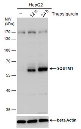
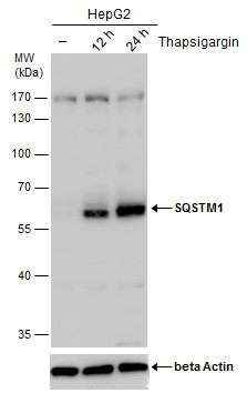
SQSTM1 antibody detects SQSTM1 protein by western blot analysis. Un-treated (-) and treated (+, Thapsigargin treatment for 12hrs and 24hrs) HepG2 whole cell extracts (30 μg) were separated by 10% SDS-PAGE, and the membrane was blotted with SQSTM1 antibody (GTX629888) diluted by 1:1000.
The ACTB was used as internal control (GTX110564, 1:50000) shown at the bottom panel.
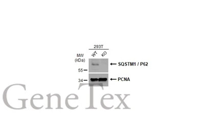
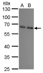
Wild-type (WT) and SQSTM1 / P62 knockout (KO) 293T cell extracts (30 μg) were separated by 10% SDS-PAGE, and the membrane was blotted with SQSTM1 / P62 antibody [GT239] (GTX629888) diluted at 1:500. The HRP-conjugated anti-mouse IgG antibody (GTX213111-01) was used to detect the primary antibody.
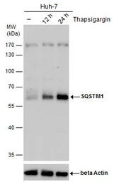
SQSTM1 antibody detects SQSTM1 protein by western blot analysis. Un-treated (-) and treated (+, Thapsigargin treatment for 12hrs and 24hrs) Huh-7 whole cell extracts (30 μg) were separated by 10% SDS-PAGE, and the membrane was blotted with SQSTM1 antibody (GTX629888) diluted by 1:1000.
The ACTB was used as internal control (GTX110564, 1:50000) shown at the bottom panel.
SQSTM1 antibody [GT239] detects SQSTM1 protein by Western blot analysis.
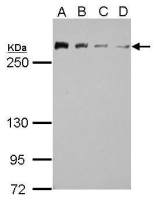
A. 30 μg A549 whole cell lysate/extract
B. 30 μg H1299 whole cell lysate/extract
10 % SDS-PAGE
SQSTM1 antibody [GT239] (GTX629888) dilution: 1:1000
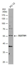
Whole cell extract (30 μg) was separated by 10% SDS-PAGE, and the membrane was blotted with SQSTM1 antibody [GT239] (GTX629888) diluted at 1:1000.
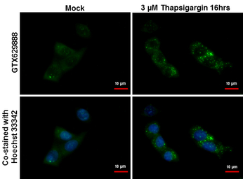
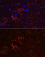
SQSTM1 antibody [GT239] detects SQSTM1 protein at autophagosome by immunofluorescent analysis.
Samples: HepG2 cells treated with 3μM thapsigargin 16 hrs (rigtht) and mock (left) were fixed in ice-cold MeOH for 10 min, permeabilize with cooled acetone for 1 min .
Green: SQSTM1 protein stained by SQSTM1 antibody [GT239] (GTX629888) diluted at 1:500.
Blue: Hoechst 33342 staining.
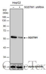
Non-transfected (–) and transfected (+) HepG2 whole cell extracts (50 μg) were separated by 10% SDS-PAGE, and the membrane was blotted with SQSTM1 antibody [GT239] (GTX629888) diluted at 1:500.
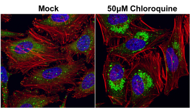
SQSTM1 antibody [GT239] detects SQSTM1 protein at autophagosome by immunofluorescent analysis.
Samples: HeLa cells mock (left) and treated with 50μM Chloroquine for 24 hr (right) were fixed in 4% PFA at RT for 15 min.
Green: SQSTM1 protein stained by SQSTM1 antibody [GT239] (GTX629888) diluted at 1:2000.
Red: phalloidin, a cytoskeleton marker, stained by phalloidin (invitrogen, A12380) diluted at 1:200.
Blue: Hoechst 33342 staining.
风险提示:丁香通仅作为第三方平台,为商家信息发布提供平台空间。用户咨询产品时请注意保护个人信息及财产安全,合理判断,谨慎选购商品,商家和用户对交易行为负责。对于医疗器械类产品,请先查证核实企业经营资质和医疗器械产品注册证情况。
 文献和实验
文献和实验Southern Blotting(Fred Hutchinson Cancer Research Cente
with Blot Wash #1: 2 x 5' at room temp (RT). 3. Wash with Blot Wash #2: 2 x 15' at 55℃. 4. Wash with Buffer 1: briefly (1') at RT. 5. (Optional; see above) Block with Buffer 2: 30' (O/N okay) Save and store at 4℃. 6. Incubate with antibody-conjugate
Detection of apoptotic process in situ using immunocytochemical and TUNEL assays
antibody (mAb) conjugated with peroxidase (anti-DNA-POD) was used. This anti-DNA-POD mAb binds to single- and double-stranded, low molecular weight DNA fragments (mono- and oligonucleosomes), showing the internucleosomal degradation of genomic DNA occuring
Phage‐Based Expression Cloning to Identify Interacting Proteins
the interacting protein. This technique leads directly to the isolation of a cDNA encoding the interacting protein, bypassing the need for labor?intensive protein purification, microsequencing, or antibody production.
 技术资料
技术资料暂无技术资料 索取技术资料

