

![Histone H2A.XS139ph (phospho Ser139) antibody [GT2311]产品图](https://img1.dxycdn.com/2022/0329/390/6616499428465020453-14.jpg)
- ¥1700 - 4000
- GeneTex

- 美国
- GTX628789
- 2026年02月10日
- WB, ICC/IF, IHC-P, IP
- Mouse
- Human, Mouse, Rat

企业认证
相关产品推荐更多 >
![ALDH7A1 antibody [C1C3]](https://img1.dxycdn.com/2022/0328/813/3734627848630300453-14.jpg!wh200)
![Caspase 8 antibody [1H11]](https://img1.dxycdn.com/2022/0328/781/3002881608403500453-14.jpg!wh200)
![INTS4 antibody [N1N2], N-term](https://img1.dxycdn.com/2022/0328/712/1489863656971300453-14.jpg!wh200)
万千商家帮你免费找货
0 人在求购买到急需产品
- 详细信息
- 文献和实验
- 技术资料
- 免疫原:
Carrier-protein conjugated synthetic peptide surrounding phospho Ser139 of human Histone H2A.X. The exact sequence is proprietary.
- 亚型:
IgG1
- 形态:
Liquid
- 保存条件:
Store as concentrated solution. Centrifuge briefly prior to opening vial. For short-term storage (1-2 weeks), store at 4ºC. For long-term storage, aliquot and store at -20ºC or below. Avoid multiple freeze-thaw cycles.
- 克隆性:
Monoclonal
- 标记物:
Unconjugated
- 适应物种:
Human, Mouse, Rat
- 保质期:
12 months from the shipping date of the product.
- 抗原来源:
Human
- 目录编号:
GTX628789
- 级别:
Primary Antibodies
- 库存:
Available
- 供应商:
GeneTex
- 宿主:
Mouse
- 应用范围:
WB, ICC/IF, IHC-P, IP
- 浓度:
0.92 mg/ml (Please refer to the vial label for the specific concentration.)
- 靶点:
Histone H2A.XS139ph (phospho Ser139)
- 抗体英文名:
Histone H2A.XS139ph (phospho Ser139) antibody [GT2311]
- 抗体名:
Histone H2A.XS139ph (phospho Ser139) 抗体 [GT2311]
- 规格:
100 μl/25 μl
| 规格: | 100 μl | 产品价格: | ¥4000.0 |
|---|---|---|---|
| 规格: | 25 μl | 产品价格: | ¥1700.0 |
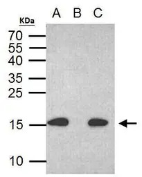
Histone H2A.X (phospho S139) antibody immunoprecipitates histone H2A.X (phospho S139) protein in IP experiments. IP Sample: 500 μg HCT116 with CPT 30 μM treatment 24 hr whole cell lysate/extract A. 30 μg HCT116 whole with CPT 30 uM treatment cell lysate/extract B. Control with 2 μg of preimmune mouse IgG C. Immunoprecipitation of histone H2A.X (phospho S139) protein by 2 μg histone H2A.X (phospho S139) antibody (GTX628789) 15% SDS-PAGE The immunoprecipitated histone H2A.X (phospho S139) protein was detected by Human histone H2A.X (phospho S139) antibody (GTX628789) diluted at 1:1000. EasyBlot anti-mouse IgG (GTX221667-01) was used as a secondary reagent.
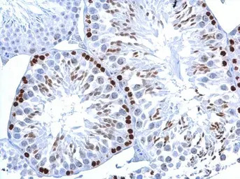
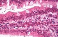
Histone H2A.XS139ph (phospho Ser139) antibody [GT2311] detects Histone H2A.XS139ph (phospho Ser139) protein at nucleus on mouse testis by immunohistochemical analysis.
Sample: Paraffin-embedded mouse testis.
Histone H2A.XS139ph (phospho Ser139) antibody [GT2311] (GTX628789) dilution: 1:500.
Antigen Retrieval: Trilogy™ (EDTA based, pH 8.0) buffer, 15min
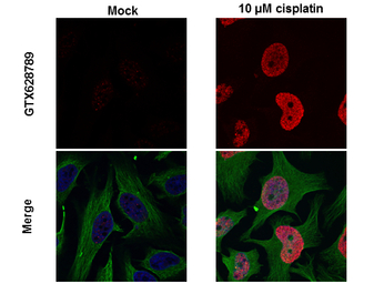
Histone H2A.X (phospho Ser139) antibody detects H2AFX protein at nuclear by confocal immunofluorescent analysis.
Sample: 10μM Cisplatin treated (right) or untreated (left) HeLa cells were fixed in 4% PFA for 15 min.
Red: H2A.X protein stained by Histone H2A.X (phospho Ser139) antibody (GTX628789) diluted at 1:500.
Green: alpha Tubulin antibody (GTX102078) diluted at 1:1000.
Blue: Hoechst 33342 staining. [Images captured by Olympus FV1000 Confocal Laser Scanning Microscope]
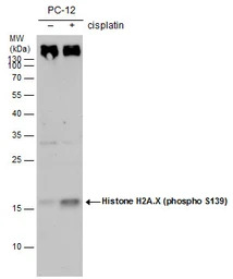
Histone H2A.X (phospho S139) antibody [GT2311] detects Histone H2A.X (phospho S139) [GT2311] protein by western blot analysis. Un-treated (-) and treated (+, 30 μM Cisplatin treatment for 24 hrs) PC-12 whole cell extracts (30 μg) were separated by 15% SDS-PAGE, and the membrane was blotted with Histone H2A.X (phospho S139) antibody [GT2311] (GTX628789) diluted by 1:500. The HRP-conjugated anti-mouse IgG antibody (GTX213111-01) was used to detect the primary antibody.
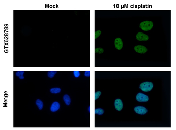
Histone H2A.X antibody detects H2AFX protein at nuclear by immunofluorescent analysis. Sample: 10μM Cisplatin treated (right) or untreated (left) HeLa cells were fixed in 4% PFA for 15 min. Green: H2AFX protein stained by Histone H2A.Xantibody (GTX628789) diluted at 1:500. Blue: Hoechst 33342 staining.
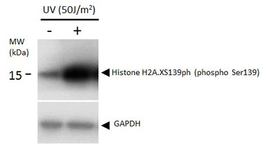
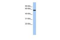
Histone H2A.XS139ph (phospho Ser139) antibody detects Histone H2A.XS139ph (phospho Ser139) protein by western blot analysis. Un-treated (-) and treated (+, 50 J/m2 UV treatment) U2OS whole cell extracts (16 μg) were separated by 12%-15% SDS-PAGE, and the membrane was blotted with Histone H2A.XS139ph (phospho Ser139) antibody (GTX628789) diluted at 1:1000. The HRP-conjugated anti-mouse IgG antibody (GTX213111-01) was used to detect the primary antibody.
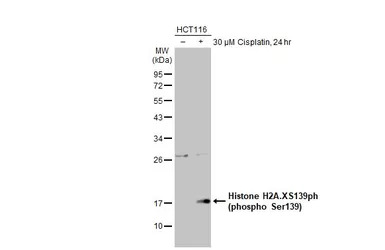
Untreated (–) and treated (+) HCT116 whole cell extracts (30 μg) were separated by 12% SDS-PAGE, and the membrane was blotted with Histone H2A.XS139ph (phospho Ser139) antibody [GT2311] (GTX628789) diluted at 1:1000. The HRP-conjugated anti-mouse IgG antibody (GTX213111-01) was used to detect the primary antibody, and the signal was developed with Trident ECL plus-Enhanced.
风险提示:丁香通仅作为第三方平台,为商家信息发布提供平台空间。用户咨询产品时请注意保护个人信息及财产安全,合理判断,谨慎选购商品,商家和用户对交易行为负责。对于医疗器械类产品,请先查证核实企业经营资质和医疗器械产品注册证情况。
 文献和实验
文献和实验Lin-Sheng Yu et al., Cancers (Basel) 2021 (PMID:34439214)
Lin BZ et al., Molecules 2020 (PMID:32784458)
Tomita T et al., Nat Commun 2021 (PMID:34135341)
Chen TY et al., Cell Death Differ 2021 (PMID:33462409)
Li G et al., J Cell Sci 2021 (PMID:33310909)
Chakraborty A et al., Nat Commun 2016 (PMID:27703167)
Honda T et al., Sci Rep 2020 (PMID:31980707)
Lin ST et al., Front Physiol 2019 (PMID:31681015)
Wang CY et al., Int J Mol Sci 2019 (PMID:31717306)
Tamanaha-Nakasone A et al., Sci Rep 2019 (PMID:31015491)
Chang CY et al., Cell Death Discov 2018 (PMID:30393570)
Wang Y et al., Neurotox Res 2014 (PMID:23996700)
En J et al., PLoS Negl Trop Dis 2017 (PMID:28783752)
Guo J et al., PLoS One 2014 (PMID:24505404)
Rodriguez A et al., Theor Biol Med Model 2015 (PMID:26385365)
Zhang Y et al., Cancer Res 2016 (PMID:27262172)
Ohba S et al., Cancer Res 2014 (PMID:25035396)
Histone H2AX Phosphorylation: A Marker for DNA Damage
) are generated that rapidly result in the phosphorylation of histone H2A variant H2AX. Because phosphorylation of H2AX at Ser 139 (γ-H2AX) is abundant, fast, and correlates well with each DSB, it is the most sensitive marker that can be used to examine the DNA
Using Phospho‐Motif Antibodies to Determine Kinase Substrates
comprising both the phosphorylated residue and the surrounding residues that determine kinase specificity, with degenerate residues taking up the remaining positions. Currently, several categories of phospho?motif antibody are commercially available
Detection of Histone H3 Phosphorylation in Cultured Cells and Tissue Sections by Immunostaining
Growth factor stimulation results in phosphorylation of histone H3 at ser 10 and this correlated with expression of immediate early genes suggesting that this phosphorylation is associated with transcriptional activation. Although Western
 技术资料
技术资料暂无技术资料 索取技术资料

