

- ¥9000
- GeneTex

- 美国
- GTX54860
- 2025年07月15日
- WB, ICC/IF, IHC-P, IHC-Fr, FACS, IP
- Rabbit
- Human, Mouse, Rat

企业认证
相关产品推荐更多 >
![PD-L1 antibody [HL2293]](https://custom.dxycdn.com/trademd/upload/pic/2013/11/12/A1383891150.jpg!small.t.1)
万千商家帮你免费找货
0 人在求购买到急需产品
- 详细信息
- 文献和实验
- 技术资料
- 免疫原:
Peptide HKLSEKLNPSVLRC, corresponding to amino acid residues 822-835 (Intracellular, C-terminus) of mouse TRPC3 (Accession : Q9QZC1).
- 亚型:
IgG
- 形态:
Liquid
- 保存条件:
Store as concentrated solution. Centrifuge briefly prior to opening vial. For short-term storage (1-2 weeks), store at 4ºC. For long-term storage, aliquot and store at -20ºC or below. Avoid multiple freeze-thaw cycles.
- 克隆性:
Polyclonal
- 标记物:
Unconjugated
- 适应物种:
Human, Mouse, Rat
- 保质期:
12 months from the shipping date of the product.
- 抗原来源:
Mouse
- 目录编号:
GTX54860
- 级别:
Primary Antibodies
- 库存:
Available
- 供应商:
GeneTex
- 宿主:
Rabbit
- 应用范围:
WB, ICC/IF, IHC-P, IHC-Fr, FACS, IP
- 浓度:
0.8 mg/ml (Please refer to the vial label for the specific concentration.)
- 靶点:
TRPC3
- 抗体英文名:
TRPC3 antibody
- 抗体名:
TRPC3 抗体
- 规格:
50 μl
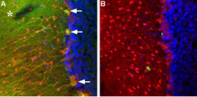
IHC-Fr analysis of rat cerebellum tissue using GTX54860 TRPC3 antibody. DAPI is used as the counterstain (blue).
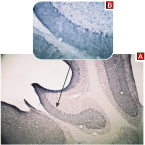
Panel A : TRPC3 (green) appears in Purkinje cells (arrows) including both soma and dendrites and as well as in the molecular layer neuropil (asterisk). Staining of the same section with mouse anti-parvalbumin (red) reveals that TRPC3 is not expressed in molecular layer interneurons.
Panel B : Pre-incubating Anti-TRPC3 with the peptide antigen
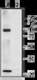
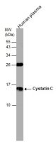
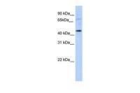
WB analysis of rat heart membrane lysate using GTX54860 TRPC3 antibody preincubated with or without immunogen peptide.
Dilution : 1:200
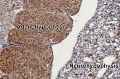
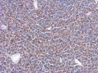
IHC-P analysis of rat pituitary gland tissue using GTX54860 TRPC3 antibody. TRPC3 is mainly expressed in the adenohypophysis (on left). Hematoxilin is used as the counterstain.
Dilution : 1:100

IHC-P analysis of mouse cerebellum tissue using GTX54860 TRPC3 antibody.
Panel A : Primary antibody
Panel B : Antibody preincubated with the control peptide antigen
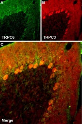
IHC-Fr analysis of rat cerebellum tissue using GTX54860 TRPC3 antibody. DAPI is used as the counterstain (blue).
Panel A : TRPC6 (green) appears in molecular layer and in Purkinje cells.
Panel B : In the same section, staining of TRPC3 (red) appears as well in both molecular layer and Purkinje cells.
Panel C : Merge images of A and B indicates extensive co-localization.
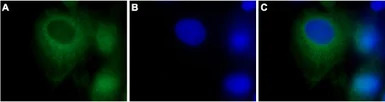
ICC/IF analysis of PFA-fixed C6 cells using GTX54860 TRPC3 antibody.
Panel A : Primary antibody
Panel B : DNA dye Hoechst 33342
Panel C : Merged images of panels A and B
Dilution : 1:500
风险提示:丁香通仅作为第三方平台,为商家信息发布提供平台空间。用户咨询产品时请注意保护个人信息及财产安全,合理判断,谨慎选购商品,商家和用户对交易行为负责。对于医疗器械类产品,请先查证核实企业经营资质和医疗器械产品注册证情况。
 文献和实验
文献和实验Ge P et al., Cell Biol Int 2018 (PMID:29570903)
Neuron:北大陈雷研究组报道胞内钙离子对 TRPC3/6 通道调控的机制
经典型瞬时受体电势通道 TRPC 是一类通透钙离子的非选择性阳离子通道1,与最早在果蝇感光系统中发现的 TRP 通道的序列相似性最高2,3,并且可以被第二信使 DAG 所激活。根据序列相似性以及通道的电生理特性,TRPC3/6/7 形成了一个亚类4。 TRPC3/6/7 通道参与多种神经活动,例如 TRPC3 在中枢神经系统高表达,参与神经生长因子 BDNF 信号转导5,同时 TRPC3 还与神经突触信号传递以及运动协调有关6,7。TRPC6 通道可以促进神经元存活以及兴奋性突触的形成
Generation of Antibody Molecules Through Antibody Engineering
been overcome to a large extent using genetic-engineering techniques to produce chimeric mouse/human and completely human antibodies. Such an approach is particularly suitable because of the domain structure of the antibody molecule ( 2 ), where functional
The importance of antibody molecules was first recognized in the 1890s, when it was shown that immunity to tetanus and diphtheria was caused by antibodies against the bacterial exotoxins (1 ). Around the same time, it was shown that antisera
 技术资料
技术资料暂无技术资料 索取技术资料

