

- ¥1700 - 4000
- GeneTex

- 美国
- GTX128839
- 2025年07月12日
- WB, ICC/IF, IHC-P, IHC-Fr, IP
- Rabbit
- Human, Mouse, Rat, Dog

企业认证
相关产品推荐更多 >
![mtPAP antibody [N1C1]](https://img1.dxycdn.com/2022/0328/279/8108060106682300453-14.jpg!wh200)
![TSPAN7 antibody [B2D]](https://img1.dxycdn.com/2022/0328/810/2155486840507700453-14.jpg!wh200)
万千商家帮你免费找货
0 人在求购买到急需产品
- 详细信息
- 文献和实验
- 技术资料
- 免疫原:
Carrier-protein conjugated synthetic peptide encompassing a sequence within the C-terminus region of human Integrin beta 1 / CD29. The exact sequence is proprietary.
- 亚型:
IgG
- 形态:
Liquid
- 保存条件:
Store as concentrated solution. Centrifuge briefly prior to opening vial. For short-term storage (1-2 weeks), store at 4ºC. For long-term storage, aliquot and store at -20ºC or below. Avoid multiple freeze-thaw cycles.
- 克隆性:
Polyclonal
- 标记物:
Unconjugated
- 适应物种:
Human, Mouse, Rat, Dog
- 保质期:
12 months from the shipping date of the product.
- 抗原来源:
Human
- 目录编号:
GTX128839
- 级别:
Primary Antibodies
- 库存:
Available
- 供应商:
GeneTex
- 宿主:
Rabbit
- 应用范围:
WB, ICC/IF, IHC-P, IHC-Fr, IP
- 浓度:
1.04 mg/ml (Please refer to the vial label for the specific concentration.)
- 靶点:
Integrin beta 1 / CD29
- 抗体英文名:
Integrin beta 1 / CD29 antibody
- 抗体名:
Integrin beta 1 / CD29 抗体
- 规格:
100 μl/25 μl
| 规格: | 100 μl | 产品价格: | ¥4000.0 |
|---|---|---|---|
| 规格: | 25 μl | 产品价格: | ¥1700.0 |
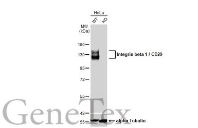
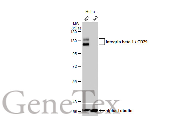
Wild-type (WT) and Integrin beta 1 / CD29 knockout (KO) HeLa cell extracts (30 μg) were separated by 7.5% SDS-PAGE, and the membrane was blotted with Integrin beta 1 / CD29 antibody (GTX128839) diluted at 1:400. The HRP-conjugated anti-rabbit IgG antibody (GTX213110-01) was used to detect the primary antibody.

Wild-type (WT) and Integrin beta 1 / CD29 knockout (KO) HeLa cell extracts (30 μg) were separated by 7.5% SDS-PAGE, and the membrane was blotted with Integrin beta 1 / CD29 antibody (GTX128839) diluted at 1:400. The HRP-conjugated anti-rabbit IgG antibody (GTX213110-01) was used to detect the primary antibody.
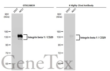
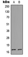
Various whole cell extracts (30 μg) were separated by 7.5% SDS-PAGE, and the membranes were blotted with Integrin beta 1 / CD29 antibody (GTX128839) diluted at 1:10000 and competitor's antibody diluted at 1:10000. The HRP-conjugated anti-rabbit IgG antibody (GTX213110-01) was used to detect the primary antibody.
*The competitor is not affiliated with GeneTex and does not endorse this product.
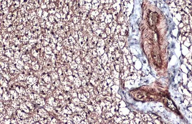
Integrin beta 1 / CD29 antibody detects Integrin beta 1 / CD29 protein at cytoplasm by immunohistochemical analysis.
Sample: Paraffin-embedded mouse brown adipocyte.
Integrin beta 1 / CD29 stained by Integrin beta 1 / CD29 antibody (GTX128839) diluted at 1:500.
Antigen Retrieval: Citrate buffer, pH 6.0, 15 min

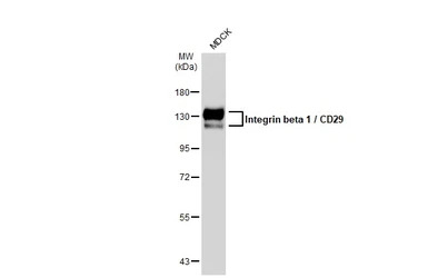
Mouse tissue extract (50 μg) was separated by 7.5% SDS-PAGE, and the membrane was blotted with Integrin beta 1 / CD29 antibody (GTX128839) diluted at 1:10000. The HRP-conjugated anti-rabbit IgG antibody (GTX213110-01) was used to detect the primary antibody.
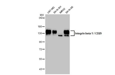
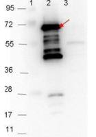
Various whole cell extracts (30 μg) were separated by 7.5% SDS-PAGE, and the membrane was blotted with Integrin beta 1 / CD29 antibody (GTX128839) diluted at 1:10000. The HRP-conjugated anti-rabbit IgG antibody (GTX213110-01) was used to detect the primary antibody.
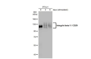
Unstimulatd and stimulatd 3T3-L1 whole cell extracts (20 μg) were separated by 7.5% SDS-PAGE, and the membrane was blotted with Integrin beta 1 / CD29 antibody (GTX128839) diluted at 1:500. The HRP-conjugated anti-rabbit IgG antibody (GTX213110-01) was used to detect the primary antibody. (The differentiation stimulated medium is composed by basal medium, 10% FBS, 50 ug/ml gentamicin, 1 nM L-glutamin, 500 uM IBMX, 1 uM dexamethasone, 2 uM rosiglitazone and 1 ug/ml insulin.)

Integrin beta 1 / CD29 antibody detects Integrin beta 1 / CD29 protein by immunohistochemical analysis.
Sample: Paraffin-embedded mouse tissues.
Integrin beta 1 / CD29 stained by Integrin beta 1 / CD29 antibody (GTX128839) diluted at 1:500.
Antigen Retrieval: Citrate buffer, pH 6.0, 15 min
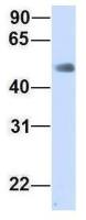
Whole cell extract (30 μg) was separated by 7.5% SDS-PAGE, and the membrane was blotted with Integrin beta 1 / CD29 antibody (GTX128839) diluted at 1:5000. The HRP-conjugated anti-rabbit IgG antibody (GTX213110-01) was used to detect the primary antibody.
风险提示:丁香通仅作为第三方平台,为商家信息发布提供平台空间。用户咨询产品时请注意保护个人信息及财产安全,合理判断,谨慎选购商品,商家和用户对交易行为负责。对于医疗器械类产品,请先查证核实企业经营资质和医疗器械产品注册证情况。
 文献和实验
文献和实验Hsiao CT et al., Oncotarget 2017 (PMID:29050309)
Wu WM et al., Int J Mol Sci 2018 (PMID:30332774)
Kao CT et al., Materials 2018;11(9)
Honig E et al., J Cell Sci 2018 (PMID:29748377)
Huang CW et al., Theranostics 2017;7(16)
Hsiao CT et al., Oncotarget 2017 (Epub)
Lu YC et al., Biochem J 2014 (PMID:24593306)
Qin N et al., Eur J Med Chem 2016 (PMID:27598237)
Zhang J et al., PLoS One 2014 (PMID:24643246)
Chung YH et al., Mol Pharm. 2014 (PMID:25153169)
Rana PS et al., Cancer Res Commun 2023 (PMID:36968231)
Chen YH et al., Cancers (Basel) 2023 (PMID:36672499)
Ngo NH et al., Front Cell Dev Biol 2022 (PMID:36120585)
M?ller M et al., Front Cell Dev Biol 2022 (PMID:35903547)
Maria J Hagelaars et al., Front Bioeng Biotechnol 2022 (PMID:35299632)
Brian A Perrino et al., Methods Mol Biol 2021 (PMID:33215378)
Ya-Hui Chen et al., Cancers (Basel) 2022 (PMID:35406599)
Ning Wang et al., J Nanobiotechnology 2022 (PMID:35236339)
Gracjana Krzysiek-Maczka et al., Am J Cancer Res 2022 (PMID:35411238)
Tan X et al., JCI Insight 2021 (PMID:34874914)
Huang CW et al., J Nanobiotechnology 2021 (PMID:34120610)
Terasaki M et al., Nutr Cancer 2021 (PMID:33590779)
Lin JW et al., Cell Death Discov 2021 (PMID:33597503)
Terasaki M et al., Cancer Genomics Proteomics 2021 (PMID:33608310)
Jin Y et al., Cell Adh Migr 2021 (PMID:33734001)
Terasaki M et al., J Clin Med 2019 (PMID:31905803)
Ou-Yang F et al., Life Sci 2019 (PMID:31639396)
Jung HJ et al., Anticancer Res 2019 (PMID:31704844)
Fujita R et al., Clin Exp Metastasis 2019 (PMID:31595388)
Xie Y et al., Anal Biochem 2019 (PMID:30981700)
Inaba H et al., PLoS One 2019 (PMID:30870452)
Li R et al., Int J Biol Macromol 2019 (PMID:30399378)
Terasaki M et al., J Nutr Biochem 2018 (PMID:30530259)
Huang CW et al., Theranostics 2017 (PMID:29109795)
CD29 分子 CD29 常用单克隆抗体或代号: 4B4;(integrin b 1,FNRb ) 主要表达细胞: 广泛分布 [AS] 分子质量(kDa)和结构: gp130血小板,GPⅡa 功 能: 与ECM粘合,细胞间粘附,结合VCAM- 1(CD106) CD29 Aka platelet GPIIa, beta-1 integrin, fibronectin receptor Positive staining
for this purpose. Grow cells in 200 mls of YPD at 30oC to an OD 600 of 1. Add formaldehyde to the 200 ml culture to a final concentration of 3.7. Fix for 1 hour at room temperature with shaking. Centrifuge cells at 3000 X g. Resuspend
Antibody cleanup In order to decrease the amount of nonspecific staining, it is often necessary to preabsorb primary and secondary antibodies to yeast cells lacking the antigen prior to use. A 1:1 mixture of fixed yeast
 技术资料
技术资料暂无技术资料 索取技术资料
