

![p63 antibody [N2C1], Internal产品图](https://img1.dxycdn.com/2022/0328/907/2607255426864200453-14.jpg)
- ¥1700 - 4000
- GeneTex

- 美国
- GTX102425
- 2025年07月10日
- WB, ICC/IF, IHC-P, IHC-Fr, IP, PLA
- Rabbit
- Human, Mouse, Rat, Dog

企业认证
相关产品推荐更多 >

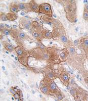
![Rap1 antibody [1D2-1C64]](https://img1.dxycdn.com/2022/0328/818/9059817428911400453-14.jpg!wh200)
![Calnexin antibody [HL1598]](https://img1.dxycdn.com/2023/0605/631/4015123267601755661-14.jpg!wh200)

万千商家帮你免费找货
0 人在求购买到急需产品
相关阅读
AJE 润色服务限时 8 折起,权威机构信任 15+ 年Cytoscape 教程来了!快速实现顶刊同款通路网络图五大应用案例:Mustang Q 膜层析应用全解析1 个小工具,一次性搞定流程图、质粒图谱和信号通路图- 详细信息
- 文献和实验
- 技术资料
- 免疫原:
Recombinant protein encompassing a sequence within the center region of human p63. The exact sequence is proprietary.
- 亚型:
IgG
- 形态:
Liquid
- 保存条件:
Store as concentrated solution. Centrifuge briefly prior to opening vial. For short-term storage (1-2 weeks), store at 4ºC. For long-term storage, aliquot and store at -20ºC or below. Avoid multiple freeze-thaw cycles.
- 克隆性:
Polyclonal
- 标记物:
Unconjugated
- 适应物种:
Human, Mouse, Rat, Dog
- 保质期:
12 months from the shipping date of the product.
- 抗原来源:
Human
- 目录编号:
GTX102425
- 级别:
Primary Antibodies
- 库存:
Available
- 供应商:
GeneTex
- 宿主:
Rabbit
- 应用范围:
WB, ICC/IF, IHC-P, IHC-Fr, IP, PLA
- 浓度:
1.58 mg/ml (Please refer to the vial label for the specific concentration.)
- 靶点:
p63
- 抗体英文名:
p63 antibody [N2C1], Internal
- 抗体名:
p63 抗体 [N2C1], Internal
- 规格:
100 μl/25 μl
| 规格: | 100 μl | 产品价格: | ¥4000.0 |
|---|---|---|---|
| 规格: | 25 μl | 产品价格: | ¥1700.0 |
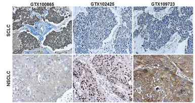
Immunohistochemical characterization of Synaptophysin (GTX100865), p63 (GTX102425) and Cytokeratin 7 (GTX109723) in human small cell lung cancer (SCLC) and non-small cell lung cancer (NSCLC) specimens.
Sample: Paraffin-embedded human SCLC (upper panel) and NSCLC (lower panel).
The section was pre-treated using heat mediated antigen retrieval with sodium citrate buffer (pH6) for 15 mins. The section was then incubated with primary antibody at 1:500 overnight at 4ºC and detected using an HRP conjugated avidin-biotin-peroxidase Complex system. DAB was used as the chromogen and counterstained with haematoxylin.
Antigen Retrieval: Citrate buffer, pH 6.0, 15 min
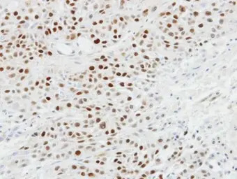
Immunohistochemical analysis of paraffin-embedded SCC4 xenograft, using p63(GTX102425) antibody at 1:100 dilution.
Antigen Retrieval: Trilogy™ (EDTA based, pH 8.0) buffer, 15min
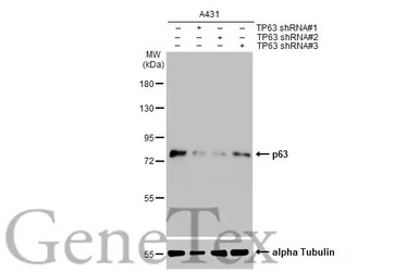
Non-transfected (–) and transfected (+) A431 whole cell extracts (30 μg) were separated by 7.5% SDS-PAGE, and the membrane was blotted with p63 antibody [N2C1], Internal (GTX102425) diluted at 1:2000. The HRP-conjugated anti-rabbit IgG antibody (GTX213110-01) was used to detect the primary antibody.
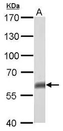
p63 antibody [N2C1], Internal detects TP63 protein by western blot analysis.
A. 50 μg mouse brain lysate/extract
7.5% SDS-PAGE
p63 antibody [N2C1], Internal (GTX102425) dilution: 1:500
The HRP-conjugated anti-rabbit IgG antibody (GTX213110-01) was used to detect the primary antibody.
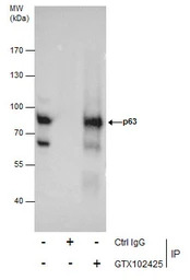
Immunoprecipitation of p63 protein from A431 whole cell extracts using 5 μg of p63 antibody [N2C1], Internal (GTX102425).
Western blot analysis was performed using p63 antibody [N2C1], Internal (GTX102425).
EasyBlot anti-Rabbit IgG (GTX221666-01) was used as a secondary reagent.
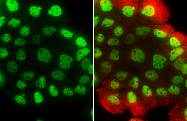
p63 antibody [N2C1], Internal detects p63 protein at nucleus by immunofluorescent analysis.
Sample: A431 cells were fixed in 4% PFA at RT for 15 min.
Green: p63 stained by p63 antibody [N2C1], Internal (GTX102425) diluted at 1:500.
Red: alpha Tubulin, stained by alpha Tubulin antibody [GT114] (GTX628802) diluted at 1:500.
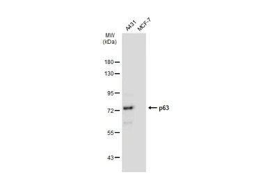
Various whole cell extracts (30 μg) were separated by 7.5% SDS-PAGE, and the membrane was blotted with p63 antibody [N2C1], Internal (GTX102425) diluted at 1:1000. The HRP-conjugated anti-rabbit IgG antibody (GTX213110-01) was used to detect the primary antibody.
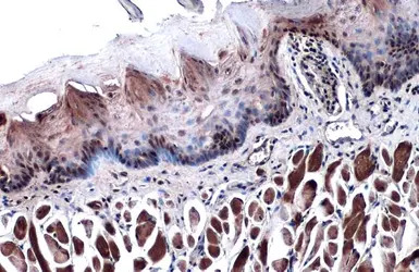
p63 antibody [N2C1], Internal detects p63 protein at nucleus by immunohistochemical analysis.
Sample: Paraffin-embedded mouse tongue.
p63 stained by p63 antibody [N2C1], Internal (GTX102425) diluted at 1:500.
Antigen Retrieval: Citrate buffer, pH 6.0, 15 min
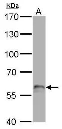
p63 antibody [N2C1], Internal detects TP63 protein by western blot analysis.
A. 50 μg rat brain lysate/extract
7.5% SDS-PAGE
p63 antibody [N2C1], Internal (GTX102425) dilution: 1:500
The HRP-conjugated anti-rabbit IgG antibody (GTX213110-01) was used to detect the primary antibody.
风险提示:丁香通仅作为第三方平台,为商家信息发布提供平台空间。用户咨询产品时请注意保护个人信息及财产安全,合理判断,谨慎选购商品,商家和用户对交易行为负责。对于医疗器械类产品,请先查证核实企业经营资质和医疗器械产品注册证情况。
 文献和实验
文献和实验人P63 酶联免疫分析(ELISA ) 试剂盒使用说明书 本试剂仅供研究使用 目的:本试剂盒用于测定人血清,血浆及相关液体样本中 P63 的 含量。 实验原理: 本试剂盒应用双抗体夹心法测定标本中人 P63 水平。用纯化的人 P63 抗体包被微孔板,制成固相抗体,往包被单抗的微孔中依次加入 P63 再与 HRP 标记的 P63 抗体结合,形成抗体 - 抗原 - 酶标抗体复合物,经过彻底洗涤后加底物 TMB 显色。 TMB 在 HRP
In the field of therapeutic recombinant proteins, monoclonal antibodies (mAbs) have achieved a rising success with more than 30 mAbs that have reached the market in the past 20 years. From a structural standpoint, one of the most important
. For most substrates, it is believed, though it has been demonstrated experimentally only for a few, that the first ubiquitin moiety is conjugated, via its C-terminal Gly76 residue, to an ε-NH2 group of an internal Lys residue. Recent findings indicate
 技术资料
技术资料暂无技术资料 索取技术资料

