

- ¥1700 - 4000
- GeneTex

- 美国
- GTX100531
- 2025年07月13日
- WB, ICC/IF, IHC-P, IP, ChIP assay
- Rabbit
- Human, Mouse

企业认证
相关产品推荐更多 >
![TIMP2 antibody [SPM356]](https://img1.dxycdn.com/2022/0328/785/3638824507411700453-14.jpg!wh200)
![CD16 antibody [2H7]](https://img1.dxycdn.com/2022/0328/589/5651429054174500453-14.jpg!wh200)
万千商家帮你免费找货
0 人在求购买到急需产品
- 详细信息
- 文献和实验
- 技术资料
- 免疫原:
Recombinant protein encompassing a sequence within the center region of human MDM2. The exact sequence is proprietary.
- 亚型:
IgG
- 形态:
Liquid
- 保存条件:
Store as concentrated solution. Centrifuge briefly prior to opening vial. For short-term storage (1-2 weeks), store at 4ºC. For long-term storage, aliquot and store at -20ºC or below. Avoid multiple freeze-thaw cycles.
- 克隆性:
Polyclonal
- 标记物:
Unconjugated
- 适应物种:
Human, Mouse
- 保质期:
12 months from the shipping date of the product.
- 抗原来源:
Human
- 目录编号:
GTX100531
- 级别:
Primary Antibodies
- 库存:
Available
- 供应商:
GeneTex
- 宿主:
Rabbit
- 应用范围:
WB, ICC/IF, IHC-P, IP, ChIP assay
- 浓度:
0.68 mg/ml (Please refer to the vial label for the specific concentration.)
- 靶点:
MDM2
- 抗体英文名:
MDM2 antibody
- 抗体名:
MDM2 抗体
- 规格:
100 μl/25 μl
| 规格: | 100 μl | 产品价格: | ¥4000.0 |
|---|---|---|---|
| 规格: | 25 μl | 产品价格: | ¥1700.0 |

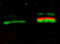
MDM2 antibody detects MDM2 protein at cytoplasm and nucleus by immunofluorescent analysis.
Sample: HepG2 cells were fixed in 4% PFA at RT for 15 min.
Green: MDM2 stained by MDM2 antibody (GTX100531) diluted at 1:500.
Red: alpha Tubulin, a cytoskeleton marker, stained by alpha Tubulin antibody [GT114] (GTX628802) diluted at 1:1000.
Scale bar= 10μm.

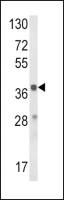
Untreated (–) and treated (+) HCT116 whole cell extract (30 μg) were separated by 7.5% SDS-PAGE, and the membrane was blotted with MDM2 antibody (GTX100531) diluted at 1:1000. The HRP-conjugated anti-rabbit IgG antibody (GTX213110-01) was used to detect the primary antibody.
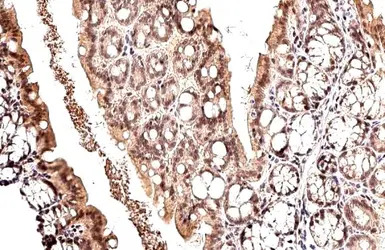
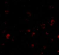
MDM2 antibody detects MDM2 protein at nucleus by immunohistochemical analysis.
Sample: Paraffin-embedded mouse colon.
MDM2 stained by MDM2 antibody (GTX100531) diluted at 1:500.
Antigen Retrieval: Citrate buffer, pH 6.0, 15 min

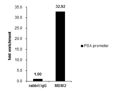
Cross-linked ChIP was performed with PC-3 chromatin extract and 5 μg of either control rabbit IgG or anti-MDM2 antibody. The precipitated DNA was detected by PCR with primer set targeting to PSA promoter.

Untreated (–) and treated (+) HepG2 whole cell extracts (30 μg) were separated by 7.5% SDS-PAGE, and the membrane was blotted with MDM2 antibody (GTX100531) diluted at 1:3000. The HRP-conjugated anti-rabbit IgG antibody (GTX213110-01) was used to detect the primary antibody.

Immunohistochemical analysis of paraffin-embedded human lung cancer, using MDM2(GTX100531) antibody at 1:500 dilution.
Antigen Retrieval: Trilogy™ (EDTA based, pH 8.0) buffer, 15min

MDM2 antibody immunoprecipitates MDM2 protein in IP experiments.
IP samples: Jurkat whole cell extract
A. 30 μg Jurkat cell whole cell extract
B. Control with 4 μg of preimmune Rabbit IgG
C. Immunoprecipitation of MDM2 protein by 4 μg MDM2 antibody (GTX100531)
7.5 % SDS-PAGE
The immunoprecipitated MDM2 protein was detected by MDM2 antibody (GTX100531) diluted at 1:500.
[EasyBlot anti-rabbit IgG (GTX221666-01) was used as a secondary reagent]
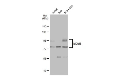
Various whole cell extracts (30 μg) were separated by 7.5% SDS-PAGE, and the membrane was blotted with MDM2 antibody (GTX100531) diluted at 1:1000. The HRP-conjugated anti-rabbit IgG antibody (GTX213110-01) was used to detect the primary antibody.
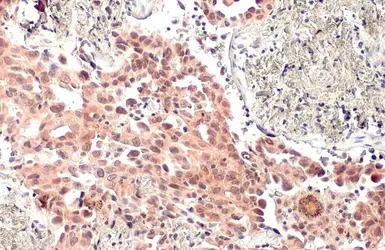
MDM2 antibody detects MDM2 protein at nucleus by immunohistochemical analysis.
Sample: Paraffin-embedded human lung cancer.
MDM2 stained by MDM2 antibody (GTX100531) diluted at 1:500.
Antigen Retrieval: Citrate buffer, pH 6.0, 15 min
风险提示:丁香通仅作为第三方平台,为商家信息发布提供平台空间。用户咨询产品时请注意保护个人信息及财产安全,合理判断,谨慎选购商品,商家和用户对交易行为负责。对于医疗器械类产品,请先查证核实企业经营资质和医疗器械产品注册证情况。
 文献和实验
文献和实验Liu Y et al., J Immunol Res 2015 (PMID:26090506)
Chai YR et al., Scand J Immunol 2014 (PMID:24965442)
Zeng M et al., Cancer Manag Res 2020 (PMID:33061576)
Zheng H et al., J Dent Res 2019 (PMID:31847675)
Soyoung I. et al., Int J Clin Exp Pathol 2018;11(4)
Tseng CW et al., Cancer Res 2018 (PMID:29599405)
Briest F et al., Neuroendocrinology 2017 (PMID:28910819)
CELL杂志04年11月份的一篇paper,介绍了单个SNP的功能学研究的方法。 点击下面链接下载 A Single Nucleotide Polymorphism in the MDM2 Promoter Attenuates the p53 Tumor Suppressor Pathway and Accelerates Tumor Formation in Humans.pdf
小红赛 我目前在做药物靶向治疗肿瘤的课题,想在肿瘤细胞水平上以P53—MDM2为靶点,研究阻断MDM2或阻止P53水解时的信号变化及对细胞生长的影响,但目前不知道在P53—MDM2这个靶点上,有什么好的药物或者抑制剂,能用于细胞水平研究的,谢谢 selleckchem01 在这个靶点 下面两个抑制剂都可以用于细胞试验~ Nutlin-3 Nutlin-3 is a MDM
Flow Cytometric Analysis of MDM2-Mediated Growth Arrest
synthesis or repair can be detected in partially denatured DNA with a BrdU-specific fluorescent antibody. Subsequent staining of transfected MDM2 with a different fluorochrome provides information about whether transfected cells make significant progression
 技术资料
技术资料暂无技术资料 索取技术资料

