大家都在搜

手机验证

询价列表

暂时没有已询价产品

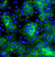
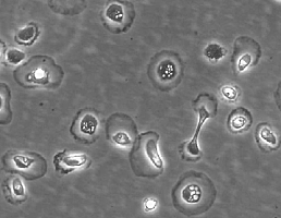
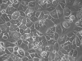
大鼠肝原代细胞

企业认证
99
biocyto
NC
Adheret 贴壁
NC
NC
肝脏 Liver
NC
大鼠
NC
梭形贴壁
否
肝 Liver
37度
6个月
存活率大于95%
10^6
| Product | Catalog no. | Amount | Storage |
| Rat Primary Hepatocytes | RHC001 | 1X10^6/vial | in liquid nitrogen |
风险提示:丁香通仅作为第三方平台,为商家信息发布提供平台空间。用户咨询产品时请注意保护个人信息及财产安全,合理判断,谨慎选购商品,商家和用户对交易行为负责。对于医疗器械类产品,请先查证核实企业经营资质和医疗器械产品注册证情况。
 询价记录
询价记录