


- 询价
- 钰博生物
- 进口/国产
- yb114300
- 2025年07月11日

企业认证
相关产品推荐更多 >
万千商家帮你免费找货
0 人在求购买到急需产品
- 详细信息
- 文献和实验
- 技术资料
- 保存条件:
-20℃
- 克隆性:
单克隆
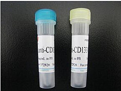
- 抗体名:
MHC Class I Qa.m2 抗体
- 规格:
0.1ml/0.2ml
MHC Class I Qa.m2
适用
小鼠
宿主
小鼠
克隆类型 (克隆位点)
单克隆 (141-16-6)
应用范围
Cytotoxicity Test (CyTox)
Pubmed 6个引用可参考
规格 0.5 mL
发货至 中国 (更改)
发货时间 29至35个工作日
产品编号 yb114300
产品细节 MHC Class I Qa.m2 抗体
免疫原 Recipient: C58/J Donor: BALB/c-H-2b Fusion Partner: P3-NSI/1-Ag4(NS-1)
克隆位点 141-16-6
亚型 IgG2b
特异性 This monoclonal antibody detects the Qa.m2 antigenic determinant on the Qa-2 Molecule. Thus, although the role of the Qa-2 antigen is unclear, antibodies to this antigen may be useful tools for separating functionally distinct subpopulations of lymphocytes and resolving pathways of differentiation. It may prove to be a useful marker in the study of B lymphocyte differentiation as well as differentiation of certain T cell functional populations.
纯化方法 Ascites
目标详细情况 MHC Class I Qa.m2 抗体
背景 The Qa region, so named because of the closely linked Qa 1-9 loci, is situated on murine chromosome 17 between the H-2D and T1a loci. These Qa loci code for cell surface alloantigens expressed on some lymphoid and myeloid cells. Their expression appears to be controlled by a gene or genes closely linked to the H-2D locus and they have been identified as being structurally related to the murine H-2 antigens. Although no functional role for Qa antigens has been determined, the restricted and differential distribution of Qa determinants on hemopoietic and lymphoid cells raises the possibility that they may not be simply differentiation markers but may play a role in hematopoetic differentiation. The Qa-2 Molecule has been identified as a two-chain structure composed of a 39,000 d heavy chain and a 12,000 d light chain which is B2 microglobulin. (The heavy chain is readily distinguished from that of H-2.) The Qa.m2 antigen has a wide representation on hematopoeitic cells. It is however, found predominantly on peripheral T cells (cytotoxic and helper T cells) but was present on only 10 % of thymus cells. Subpopulations of B cells (approximately 25 % of all B cells) and bone marrow cells (15-20 %) are also reactive. Qa.m2 was also detected on megakaryocyte colony-forming cells (MEG-CFC) but not on granulocyte- macrophage, erythroid or eosinophil colonies. In addition, the Qa.m2 is a marker of T cell differentiation, as it is selectively expressed on functional T cells. The control of Qa-2 antigen expression and of other Qa antigens by the H-2 region may reflect a control mechanism of functional T cell differentiation and ontogeny in developing T lymphocytes.
基因ID 110557
NCBI登录号 NP_997531
UniProt P79568
研究领域 Major Histocompatibility Complex (MHC), Immunology
使用细节 MHC Class I Qa.m2 抗体
应用备注 Cytotoxicity Analysis. Flow Cytometry.
Other applications not tested.
Optimal dilutions are dependent on conditions and should be determined by the user.
实验流程 RECOMMENDED METHOD FOR DEPLETING A CELL POPULATION OF Qa. m2 POSITIVELYMPHOCYTES1. Prepare a cell suspension from the appropriate tissue in Cytotoxicity Medium orequivalent. Remove red cells and dead cells (where necessary) by purification of viablelymphocytes on Lympholyte®-M density cell separation medium . After washing adjust thecell concentration to 1 x 10e7 cells per ml in Cytotoxicity Medium. 2. Add the antibody to a final concentration 1: 20 and mix. Alternately, pellet the cells andresuspend in antibody diluted 1: 20 in Cytotoxicity Medium. 3. Incubate for 60 minutes at 4°C. 4. Centrifuge to pellet the cells and discard the supernatant. 5. Resuspend to the original volume in Low-Tox®-M Rabbit Complement3, diluted to theappropriate concentration in Cytotoxicity Medium. (Recommended concentration included
限制 仅限研究用
贮存及处理 MHC Class I Qa.m2 抗体
状态 Liquid
溶解方式 Restore with 0.5 mL of distilled water.
注意事项 Use of Sodium Azide as a preservative will substantially inhibe the activity of Horseradish peroxidase (HRP).
注意事项 Avoid repeated freezing and thawing. This product is photosensitive and should be protected from light. Should it contain a precipitate we recommend microcentrifugation before use.
储存条件 -20 °C
储存方法 Prior to reconstitution store the antibody at 2-8 °C. Following reconstitution store undiluted at 2-8 °C for one month or at -20 °C for longer.
风险提示:丁香通仅作为第三方平台,为商家信息发布提供平台空间。用户咨询产品时请注意保护个人信息及财产安全,合理判断,谨慎选购商品,商家和用户对交易行为负责。对于医疗器械类产品,请先查证核实企业经营资质和医疗器械产品注册证情况。
 文献和实验
文献和实验MHC I 类分子在 CD8+细胞免疫系统中起到抗原呈递和识别作用。因其特异性,MHC-多肽复合物在免疫治疗中起着重要作用。虽然 MHC-多肽复合物能够被 TCR 特异性识别,但大多数天然 TCR 受体不仅稳定性低,与 MHC-多肽复合物的亲和力也低。为了解决上述两个不足,以肿瘤抗原表位为靶点,制备亲和力高,且可溶性强的 TCR 样抗体成为替代野生型 TCR 受体的最佳选择。该类 TCR 样抗体作为一种新的免疫分子,起着识别并杀灭肿瘤细胞的作用。1、以 T 细胞表位为靶标的免疫治疗自身免
1、流式细胞仪上的FL2-W FL2-A FL2-H 分别是做什么的? FL2-W是只检测荧光的脉冲宽度, FL2-H是指脉冲高度。通常在做细胞周期分析时应用,用于去除粘连细胞。 2、流式同型对照怎样选择? 同型对照(Isotype Control):使用与一抗相同种属来源、相同亚型、相同剂量和相同的免疫球蛋白及亚型的免疫球蛋白,用于消除由于抗体非特异性结合到细胞表面而产生的背景染色。如果一抗是多抗,可以用an normal serum(与一抗相同的正常
免疫细胞膜分子(一):主要组织相容性抗原-- MHC基因结构
(histocompatibility antigen)或移植抗原(transplantation antigen)。机体内与排斥反应有关的抗原系统多达20种以上,其中能引起强而迅速排斥反应者称为主要组织相容性抗原,其编码基因是一组紧密连锁的基因群,称为主要组织相容性复合体(major histocompatibility complex,MHC)。现已证明,控制机体免疫应答能力与调节功能的基因(immune uesponse gene,Ir gene )也存在于MHC内。因此,MHC不仅与移植排斥反应有关,也广泛参与免疫
 技术资料
技术资料暂无技术资料 索取技术资料

