

- ¥3870
- biorbyt

- orb10487
- 英国
- 2025年12月09日
- ICC, IF, IHC-P, WB
- Rabbit
- Guinea pig, Human, Rat

企业认证

研选同类产品更多 >


万千商家帮你免费找货
0 人在求购买到急需产品
- 详细信息
- 用户评价
- 文献和实验
- 技术资料
- 抗体名:
CXCR1 antibody
- 抗体英文名:
CXCR1 antibody
- 靶点:
CXCR1
- 浓度:
- 100 μg (in 200 μl): 0.5 mg/ml- 200 μg (in 400 μl): 0.5 mg/ml
- 应用范围:
ICC, IF, IHC-P, WB
- 宿主:
Rabbit
- 适应物种:
Guinea pig, Human, Rat
- 保质期:
6-12个月
- 抗原来源:
详询
- 目录编号:
orb10487
- 级别:
科研
- 库存:
99
- 供应商:
Biorbyt
- 标记物:
Unconjugated
- 克隆性:
Polyclonal
- 保存条件:
低温
- 形态:
10 mM PBS, 0.02% sodium azide
- 亚型:
IgG
- 免疫原:
KLH conjugated synthetic peptide derived from human CXCR1. Please contact us for the exact immunogen sequence. The peptide is available as orb12890.
- 规格:
100 ug
产品名:CXCR1 antibody
货号:orb10487
产品中文名:CXCR1 抗体
产品详情链接:https://www.biorbyt.com.cn/cxcr1-antibody-orb10487.html
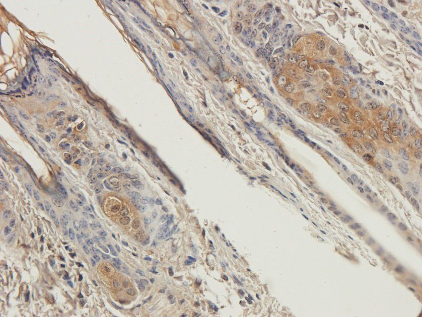
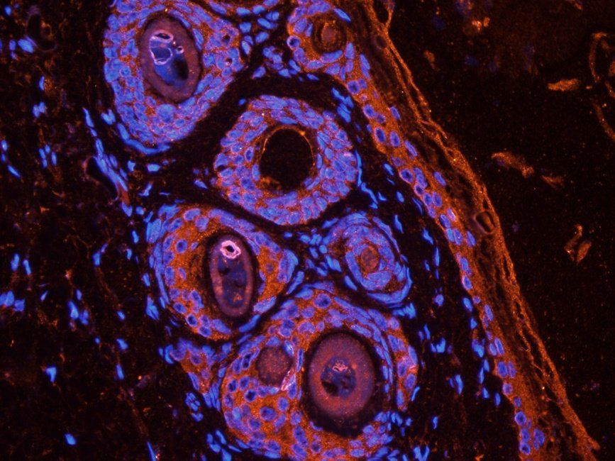
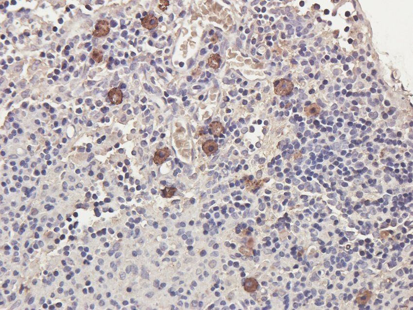
IHC-P image of rat lymph node tissue using anti-CXCR1 (2.5 ug/ml)

武汉博欧特生物科技有限公司( www.wuhanbio.com )为英国品牌Biorbyt( www.biorbyt.com )中国办事处,即biorbyt中国总代理,即biorbyt总代。负责biorbyt品牌产品在中 国市场的推广、销售及售后服务。为您提供各种biorbyt原装进口产品,主推四 大类:
一. 抗体(antibody):24万种的一抗。 可应用于Western blot,IP,IHC,IF,FACS,ELISA等实验。
二. 蛋白(protien):3.6万种的高纯度蛋白 。
三. elisa试剂盒(elisa kit):可以提 供超过1.5万种的ELISA试剂盒,包括人源,大鼠源,小鼠源等。
四. 小分子(Small Molecules):超过 1.5万种的生物活性小分子,主要包括:拮抗剂、抑制剂、抗生素、天然产物。
所有biorbyt品牌的抗体,蛋白,elisa试剂 盒,小分子等产品国际航空低温运输,货期短,多数产品货期在1~两周,快捷 便利。
我们为研究者们提供免费的专业的售前售中 售后服务,为您的实验保驾护航。
公司对外诚接特色技术外包服务如下:
重组蛋白表达与纯化服务、单克隆抗体定制 服务、多克隆抗体定制服务、免疫学检测服务、动物实验技术服务。我们为客户 提供详实的生物信息咨询和全方位的可行性分析方案,为您的科研项目保驾护航 。更多详情请技术咨询027-87663101 / 15827569716。
风险提示:丁香通仅作为第三方平台,为商家信息发布提供平台空间。用户咨询产品时请注意保护个人信息及财产安全,合理判断,谨慎选购商品,商家和用户对交易行为负责。对于医疗器械类产品,请先查证核实企业经营资质和医疗器械产品注册证情况。
 用户评价
用户评价 暂无用户评价
 文献和实验
文献和实验Pharmacology, Biodistribution, and Efficacy of GPCR-Based Pepducins in Disease Models
against a wide variety of GPCRs including the protease-activated receptors (PAR1, 2, 4), the chemokine receptors (CXCR1, 2, 4), the sphingosine-1-phosphate receptor (S1P3), the adrenergic receptor (ADRA1B), and have the potential to help reveal the functions
Examples of Involvement in Disease CCR5 provides a signal for microbial induced production of IL-12 by CD8+ dendritic cells Regulation of tyrosine kinase activation and granule release through -arrestin by CXCR1
让肿瘤变「热」!宋尔卫院士团队Nat Immunol发文报道肿瘤免疫治疗新策略
(2019).9. L. Jin et al., CXCR1- or CXCR2-modified CAR T cells co-opt IL-8 for maximal antitumor efficacy in solid tumors. Nat Commun 10, 4016 (2019).10. L. Zhao, Y. J. Cao, Engineered T Cell Therapy for Cancer in the Clinic. Front Immunol 10, 2250 (2019


