

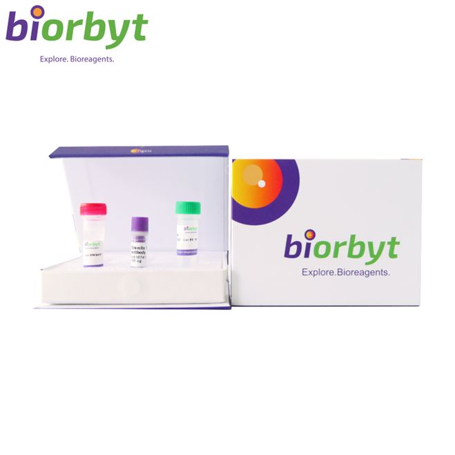
- ¥8775
- Biorbyt

- orb2644690
- 英国
- 2026年08月07日
- IHC-P
- Mouse
- Human

企业认证
相关产品推荐更多 >
万千商家帮你免费找货
0 人在求购买到急需产品
相关阅读
细胞污染识别与处理全攻略:5 种常见类型+关键误区Cytoscape 教程来了!快速实现顶刊同款通路网络图五大应用案例:Mustang Q 膜层析应用全解析1 个小工具,一次性搞定流程图、质粒图谱和信号通路图- 详细信息
- 文献和实验
- 技术资料
- 抗体名:
Recombinant PHH3 Antibody / Histone H3 (pSer10) 抗体
- 抗体英文名:
Recombinant PHH3 Antibody / Histone H3 (pSer10)
- 应用范围:
IHC-P
- 宿主:
Mouse
- 适应物种:
Human
- 保质期:
12 months from date of receipt.
- 抗原来源:
详询
- 目录编号:
orb2644690
- 级别:
科研
- 库存:
99
- 供应商:
Biorbyt
- 标记物:
Unconjugated
- 克隆性:
Recombinant
- 保存条件:
详询
- 亚型:
Mouse IgG2a, kappa
- 免疫原:
详询
- 规格:
100 ug
产品子类型: Primary Antibody
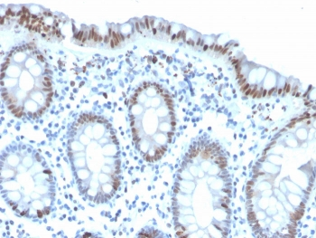
描述: Phosphohistone H3 (PHH3) is a marker specific for cells undergoing mitosis. Serine 10 of Histone H3 is phosphorylated in association with mitotic chromatin condensation in late G2 and M phase of the cell cycle and thus, PHH3 can distinguish mitosis from apoptotic nuclei. The range of percentage PHH3 positive tumor nuclei was from 0.0 to 6.6% (median value 0.8%). Increased expression of PHH3 was significantly associated with tumor thickness (p = 0.031), presence of tumor ulceration (p =0.041) and tumor necrosis (p =0.027), but not with Clarks level of invasion. High levels of PHH3 was associated with increased mitotic count (p = 0.003) and high Ki-67 expression (p = 0.002). For central nervous system tumors, melanoma, soft tissue tumors, GIST, etc., PHH3 mAb is helpful for tumor pathological classification and prognosis.
克隆号: PHH3/471R
应用稀释比例: Immunohistochemistry (FFPE): 1-2ug/ml for 30 minutes at RT
应用说明: Optimal dilution of the recombinant PHH3 antibody should be determined by the researcher.
免疫原: A synthetic peptide corresponding to the amino acids surrounding phospho S10 were used as the immunogen for the recombinant PHH3 antibody.
防腐剂: 1 mg/ml in 1X PBS; rAlbumin free, sodium azide free
纯化方法: Protein G affinity chromatography
研究领域: Epigenetics & Chromatin
UniProt ID: P68431
保存条件: Maintain refrigerated at 2-8°C for up to 2 weeks. For long term storage store at -20°C in small aliquots to prevent freeze-thaw cycles.
备注: For research use only.
风险提示:丁香通仅作为第三方平台,为商家信息发布提供平台空间。用户咨询产品时请注意保护个人信息及财产安全,合理判断,谨慎选购商品,商家和用户对交易行为负责。对于医疗器械类产品,请先查证核实企业经营资质和医疗器械产品注册证情况。
 文献和实验
文献和实验Detection of Histone H3 Phosphorylation in Cultured Cells and Tissue Sections by Immunostaining
. The protocol described here allows the detection of phosphorylated histones in tissue-cultured cells and tissue sections by fluorescent or bright-field immunostaining analysis. Here we used a serine 10 specific P-histone H3 antibody to determine
3X Flag Peptide) to each pelleted bead/complex, incubate 30-60 minutes at 4°C with gentle shaking;Centrifuge the resin for 30 seconds at 10,000 RPM, transfer the supernatants (which should now contain the ChIP/Flag eluted complexes) to fresh tubes
by vortexing every 10 min. Stop reaction by adding 15 µL of 4x sample buffer. Heat on 95°C block for 5 min. (Can be stored at 4°C overnight) Spin down beads and load supernatant on a 12% SDS acrylamide gel. Run off the dye front (175 volts x
 技术资料
技术资料暂无技术资料 索取技术资料

