

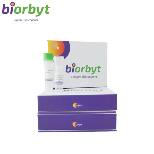
- ¥5499
- Biorbyt

- orb1291730
- 英国
- 2026年07月31日
- ELISA, FC, IHC, WB
- Rabbit
- Human, Mouse, Rat

企业认证
相关产品推荐更多 >
CTDSP2 Rabbit Polyclonal Antibody (Biotin) 抗体,orb2109835,Biorbyt
¥0
IgG (Immunoglobulin Gamma Heavy Chain) (B-Cell Marker) Antibody - With BSA and Azide 抗体,orb1462396,Biorbyt
¥6084
SLC14A1 Rabbit Polyclonal Antibody 抗体,orb101768,Biorbyt
¥3159
ANXA10 Antibody 抗体,orb1338071,Biorbyt
¥7488
BRD1 Antibody (N-term) 抗体,orb32103,Biorbyt
¥2106
万千商家帮你免费找货
0 人在求购买到急需产品
相关阅读
源叶现货试剂/标准品,库存足,周末发货Cytoscape 教程来了!快速实现顶刊同款通路网络图五大应用案例:Mustang Q 膜层析应用全解析1 个小工具,一次性搞定流程图、质粒图谱和信号通路图- 详细信息
- 文献和实验
- 技术资料
- 抗体名:
MST3/STK24 Rabbit Polyclonal Antibody 抗体
- 抗体英文名:
MST3/STK24 Rabbit Polyclonal Antibody
- 靶点:
Serine/threonine-protein kinase 24
- 浓度:
Adding 0.2 ml of distilled water will yield a concentration of 500 μg/ml.
- 应用范围:
ELISA, FC, IHC, WB
- 宿主:
Rabbit
- 适应物种:
Human, Mouse, Rat
- 保质期:
12 months from date of receipt.
- 抗原来源:
详询
- 目录编号:
orb1291730
- 级别:
科研
- 库存:
99
- 供应商:
Biorbyt
- 标记物:
Unconjugated
- 克隆性:
Polyclonal
- 保存条件:
详询
- 形态:
Lyophilized
- 亚型:
Rabbit IgG
- 免疫原:
详询
- 规格:
100 ug
产品子类型: Primary Antibody
描述: Anti-MST3/STK24 Antibody. Tested in ELISA, Flow Cytometry, IHC, WB applications. This antibody reacts with Human, Mouse, Rat.
别名: MST 3; MST3; MST3B; STE20; STE20 like kinase MST3; STK24; STK3
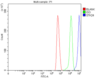
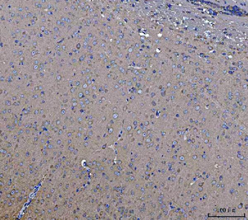
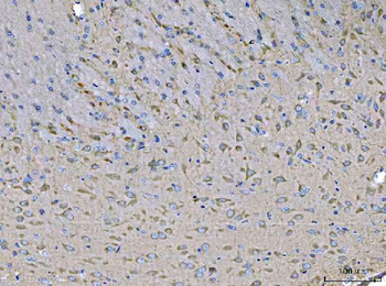
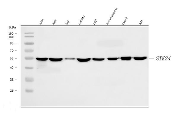
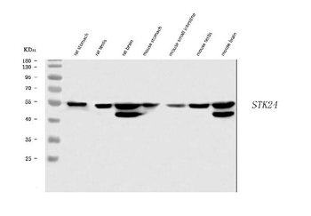
应用稀释比例: Western blot, 0.1-0.25 μg/ml, Human, Mouse, Rat Immunohistochemistry(Paraffin-embedded Section), 2-5 μg/ml, Mouse, Rat Flow Cytometry (Fixed), 1-3 μg/1x10^6 cells, Human ELISA, 0.1-0.5 μg/ml
免疫原: E.coli-derived human MST3/STK24 recombinant protein (Position: M1-Y433).
交叉反应: No cross-reactivity with other proteins.
分子量: 55 kDa
防腐剂: Each vial contains 4 mg Trehalose, 0.9 mg NaCl, 0.2 mg Na2HPO4.
纯化方法: Immunogen affinity purified.
复溶/重构: Adding 0.2 ml of distilled water will yield a concentration of 500 μg/ml.
研究领域: Cell Biology, Signal Transduction
UniProt ID: Q9Y6E0
保存条件: Maintain refrigerated at 2-8°C for up to 2 weeks. For long term storage store at -20°C in small aliquots to prevent freeze-thaw cycles.
备注: For research use only.
风险提示:丁香通仅作为第三方平台,为商家信息发布提供平台空间。用户咨询产品时请注意保护个人信息及财产安全,合理判断,谨慎选购商品,商家和用户对交易行为负责。对于医疗器械类产品,请先查证核实企业经营资质和医疗器械产品注册证情况。
 文献和实验
文献和实验Polyclonal Antibody Production
Zealand White Rabbits and have them delivered to the ARC. Don’t put all your eggs in one basket only using one rabbit. Let them get accustomed to there home there for two weeks before the first pre-bleed. Pre-bleed
Polyclonal Antibody Production
Polyclonal Antibody Production Very useful for rapid and simple generation of antibodies for western blots, ELISA assays, and immunoprecipitation. Rabbit Immunization Initial Preparation Your antigen
Generation and Selection of Rabbit Antibody Libraries by Phage Display
The rabbit antibody repertoire is an exceptional source for both polyclonal antibodies (pAbs) and monoclonal antibodies (mAbs) that combine high specificity with high avidity and affinity, respectively. In contrast to rabbit pAbs
 技术资料
技术资料暂无技术资料 索取技术资料

