

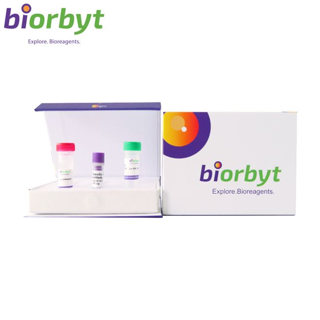
- ¥3159 - 4797
- Biorbyt

- orb763954
- 英国
- 2026年07月24日
- ELISA, WB
- Human, Mouse, Rat

企业认证
相关产品推荐更多 >
万千商家帮你免费找货
0 人在求购买到急需产品
相关阅读
AJE 润色服务限时 8 折起,权威机构信任 15+ 年Cytoscape 教程来了!快速实现顶刊同款通路网络图五大应用案例:Mustang Q 膜层析应用全解析1 个小工具,一次性搞定流程图、质粒图谱和信号通路图- 详细信息
- 文献和实验
- 技术资料
- 抗体名:
Histone H3 (Mono Methyl Lys10) rabbit pAb Antibody 抗体
- 抗体英文名:
Histone H3 (Mono Methyl Lys10) rabbit pAb Antibody
- 浓度:
1 mg/ml
- 应用范围:
ELISA, WB
- 适应物种:
Human, Mouse, Rat
- 保质期:
12 months from date of receipt.
- 抗原来源:
详询
- 目录编号:
orb763954
- 级别:
科研
- 库存:
99
- 供应商:
Biorbyt
- 标记物:
Unconjugated
- 克隆性:
Polyclonal
- 保存条件:
详询
- 亚型:
IgG
- 免疫原:
详询
- 规格:
50 ul/100 ul
| 规格: | 50 ul | 产品价格: | ¥3159.0 |
|---|---|---|---|
| 规格: | 100 ul | 产品价格: | ¥4797.0 |
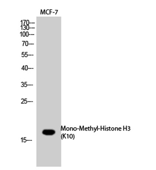
描述: Rabbit polyclonal antibody to Histone H3 (Mono-Methyl-Lys10)
别名: Anti-H3K10ME1 antibody, anti-HIST1H3A antibody, anti-H3FA antibody, anti-HIST1H3B antibody, anti-H3FL antibody, anti-HIST1H3C antibody, anti-H3FC antibody, anti-HIST1H3D antibody, anti-H3FB antibody, anti-HIST1H3E antibody, anti-H3FD antibody, anti-HIST1H3F antibody, anti-H3FI antibody, anti-HIST1H3G antibody, anti-H3FH antibody, anti-HIST1H3H antibody, anti-H3FK antibody, anti-HIST1H3I antibody, anti-H3FF antibody, anti-HIST1H3J antibody, anti-H3FJ antibody, anti-Histone H3.1 antibody, anti-Histone H3.1 antibody, anti-His antibody
应用稀释比例: Western Blot: 1/500 - 1/2000. ELISA: 1/20000. Not yet tested in other applications.
免疫原: Synthesized peptide derived from human Histone H3 around the mono-methylation site of K10.
分子量: 17
防腐剂: PBS with 0.02% sodium azide and 50% glycerol pH 7.4.
纯化方法: The antibody was affinity-purified from rabbit antiserum by affinity-chromatography using epitope-specific immunogen.
来源: Rabbit
研究领域: Epigenetics & Chromatin
UniProt ID: Q6NXT2, Q71DI3, P68431, P84243
保存条件: Maintain refrigerated at 2-8°C for up to 2 weeks. For long term storage store at -20°C in small aliquots to prevent freeze-thaw cycles.
备注: For research use only.
风险提示:丁香通仅作为第三方平台,为商家信息发布提供平台空间。用户咨询产品时请注意保护个人信息及财产安全,合理判断,谨慎选购商品,商家和用户对交易行为负责。对于医疗器械类产品,请先查证核实企业经营资质和医疗器械产品注册证情况。
 文献和实验
文献和实验Detection of Histone H3 Phosphorylation in Cultured Cells and Tissue Sections by Immunostaining
. The protocol described here allows the detection of phosphorylated histones in tissue-cultured cells and tissue sections by fluorescent or bright-field immunostaining analysis. Here we used a serine 10 specific P-histone H3 antibody to determine
in detail here. The first protocol is essentially a dual-pulldown assay employing Flag-tagged DNMT3A or antibody of choice for an endogenous protein and 5' biotin linked antisense RNA (Figure 1a), while the second protocol is a triple pulldown assay which essentially expands
RUVKUN Antibody Staining Protocol revised 10/29/90
RUVKUN Antibody Staining Protocol revised 10/29/90 Fixation: 1. C. elegans N2 grown in liquid or plates. Harvest worms. Wash for 2 hours at room temp. in 1X M9 or PBS. 2. Fix in 5 ml of FRESH THAT DAY 1% paraformaldehyde:
 技术资料
技术资料暂无技术资料 索取技术资料

