


- ¥8580
- biorbyt

- orb148177
- 英国
- 2025年12月04日
- AM, ICC, IF, IHC, IP, WB
- Mouse
- Human, Mouse, Rat

企业认证
相关产品推荐更多 >


万千商家帮你免费找货
0 人在求购买到急需产品
- 详细信息
- 文献和实验
- 技术资料
- 抗体名:
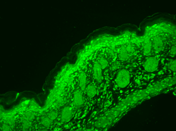
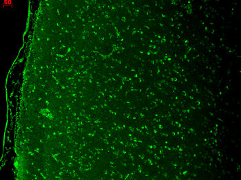
Cav1.3 Antibody (APC)抗体
- 抗体英文名:
Cav1.3 Antibody (APC)
- 靶点:
Cav1.3
- 浓度:
1 mg/ml
- 应用范围:
AM, ICC, IF, IHC, IP, WB
- 宿主:
Mouse
- 适应物种:
Human, Mouse, Rat
- 保质期:
6-12个月
- 抗原来源:
详询
- 目录编号:
orb148177
- 级别:
科研级
- 库存:
88
- 供应商:
biorbyt
- 标记物:
APC
- 克隆性:
Recombinant
- 亚型:
IgG2a Kappa
- 免疫原:
Fusion protein amino acids 859-875 of rat Cav1.3
- 规格:
100 ug
别名:Voltage-dependent L-type calcium channel subunit alpha-1D, Calcium channel, L type, alpha-1 polypeptide isoform 2, Voltage-gated calcium channel subunit alpha Cav1.3, CACNA1D, CACH3, CACN4, CACNL1A2, CCHL1A2, alpha-1 polypeptide, CAC1D_HUMAN, Cacna1d, Calcium channel, Calcium channel L type alpha 1 polypeptide isoform 2, Calcium channel neuroendocrine/brain type alpha 1 subunit, Calcium channel voltage dependent L type alpha 1D subunit, isoform 2, L type, Voltage dependent L type calcium channel subunit alpha 1D, Voltage gated calcium channel alpha 1 subunit, Voltage gated calcium channel alpha subunit Cav1.3
免疫原:Fusion protein amino acids 859-875 of rat Cav1.3
克隆性:S48
分子量:250kDa
应用稀释比例:WB (1:1000), IHC (1:1000), ICC/IF (1:100)
应用注释:1 µg/ml of SMC-301 was sufficient for detection of Cav1.3 in 10 µg of rat brain lysate by colorimetric immunoblot analysis using Goat anti-mouse IgG:HRP as the secondary antibody.
防腐剂:95.46mM Phosphate, 2.48mM MES and 2mM EDTA
纯化:Protein G Purified
保存说明:Conjugated antibodies should be stored according to the product label
NCBI:NP_058994.1
Entrez:29716
UniProt ID:P27732
Note:For research use only.

风险提示:丁香通仅作为第三方平台,为商家信息发布提供平台空间。用户咨询产品时请注意保护个人信息及财产安全,合理判断,谨慎选购商品,商家和用户对交易行为负责。对于医疗器械类产品,请先查证核实企业经营资质和医疗器械产品注册证情况。
 文献和实验
文献和实验抗体的概念 抗体(antibody, Ab),也叫免疫球蛋白(immunoglobulin, Ig),是血液和组织液中的一类糖蛋白,由B细胞接受抗原刺激后增殖分化成的浆细胞产生,能与相应抗原特异性地结合,是介导体液免疫的重要效应分子。1968年和1972年世界卫生组织和国际免疫学联合会的专门委员会先后决定,将具有抗体活性或化学结构与抗体相似的球蛋白统称为免疫球蛋白。后来研究证实,Ig和Ab在结构及功能上完全一致,因此可认为二者的概念等同。Ig可分为分泌型和跨模型,前者主要存在于血液和组织液中
v468 Chapter 6 细胞内结肠腺瘤样息肉蛋白核定位以及细胞质检测
, and has characterized pathways for the trafficking of APC both into and out of the nucleus.Antibody specificity is an important factor in the determination of APC localization, and in this chapterwe outline a strategy for the unambiguous detection of APC using
Biosynthetic labeling (Sefton Lab) Biotinylation of Antibody (Contributed by Nanci Donacki) 125 I Labeling of Protein using ICl (ScienceXchange) Protein (Antibody) Fluorescent Labeling (Mario Roederer) Gives detailed
 技术资料
技术资料暂无技术资料 索取技术资料

