


- ¥5425
- other
- 009
- 中国
- 2026年04月24日

企业认证


万千商家帮你免费找货
0 人在求购买到急需产品
- 详细信息
- 文献和实验
- 技术资料
- 品系:
原代
- 细胞类型:
原代细胞
- 肿瘤类型:
无
- 供应商:
上海然其生物科技有限公司
- 库存:
现货
- 英文名:
Human pulmonary artery endothelial cells
- 生长状态:
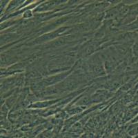
长梭状细胞,贴壁培养
- 年限:
无
- 运输方式:
常温/干冰
- 器官来源:
人
- 是否是肿瘤细胞:
否
- 细胞形态:
长梭状细胞
- 免疫类型:
平滑肌肌动蛋白(α-SMA)免疫荧光染色为阳性
- 物种来源:
人
- 相关疾病:
无
- 组织来源:
人的正常肺动脉组织
细胞详述
肺动脉起于右心室,在主动脉之前向左上后方斜行,在主动脉弓下方分为左、右肺动脉,经肺门入肺。由于肺动脉连接着输送静脉血的右心室,所以,肺动脉虽然是动脉,但是它却输送静脉血。血管平滑肌细胞互相连接,形成管状结构;在功能上可以通产生连续收缩,使器官对抗所加负荷而保持原有的形状。该细胞所表达的钙通道表面表达的ICAM-1和VCAM-1,参与血管壁炎症反应。体外培养的肺大动脉平滑肌细胞呈梭形、星形或不规则形。
细胞特性
1) 组织来源于人的正常肺动脉组织。
2) 细胞鉴定:平滑肌肌动蛋白(α-SMA)免疫荧光染色为阳性。
3) 经鉴定细胞纯度高于90%。
4) 不含有 HIV-1、 HBV、HCV、支原体、细菌、酵母和真菌。
5) 细胞生长方式:长梭状细胞,贴壁培养。
产品的运输和保存
视天气状况和运输距离远近,公司与客户协商后选择下述方式中的一种进行。
1)1mL冻存细胞悬液装于1.8ml的冻存管中,置于装满干冰的泡沫保温盒中进行运输;收到细胞后请尽快解冻复苏细胞进行培养,如无法立刻进行复苏操作,冻存细胞可在-80℃的条件下保存1个月。
2)T-25培养瓶充满完全培养基后进行常温运输;收到细胞后请镜下观察细胞生长状态,如铺瓶率超过85%请立即进行传代操作,如悬浮的细胞较多,请将培养瓶至于培养箱中静置过夜以帮助未死亡的悬浮细胞能够再次贴壁。
产品使用
1)本产品仅能用于科研
2)本产品未通过直接用于活体动物和人的审核
3)本产品未通过用于活体诊断的审核
Cell Details
Cell Characteristics
Shipping and Storage
Product Usage
风险提示:丁香通仅作为第三方平台,为商家信息发布提供平台空间。用户咨询产品时请注意保护个人信息及财产安全,合理判断,谨慎选购商品,商家和用户对交易行为负责。对于医疗器械类产品,请先查证核实企业经营资质和医疗器械产品注册证情况。
 文献和实验
文献和实验1、传代前24h先将组织块去除:等到细胞覆盖瓶底80%以上后进行组织块去除操作。可用弯头玻璃吸管将组织块轻轻挑起吸出,确保无组织块留下,以防坏死污染。2、胰酶消化:倒掉旧培养液,用D-HANKs液清洗两边,加入已预热(37度)10min的0.25%胰酶消化液2ml,2-3min后镜下观察,发现胞质收缩,细胞间隙增大,立即用含胎牛血清的培养液终止消化。3、弯头吸管吹打细胞:吹打一定时间后到镜下观察,在没吹打起细胞区域继续吹打,动作要柔和。4、离心:2000转5min。倒掉上清液,加入培养液,吹
实验步骤(一)、取SD 大鼠一只,实验时断椎处死。(二)、在无菌条件下快速自肛门上2 cm 取结肠10 cm 左右,生理盐水中反复灌肠冲洗。(三)、移入含300 U/ml青霉素、300 U/ml 链霉素的HepesRinger缓冲液中浸泡,肠段内外各15 min。(四)、将经抗生素浸泡过的肠段移入含Hepes Ringer缓冲液的塑料培养板中(培养板预先铺上705胶),在体视显微镜下沿肠系膜对侧纵行切开肠段,皮内针固定好四角,仔细地刮除粘膜层,翻转至外侧,小心地撕去肠系膜,再仔细地刮去浆膜
1、麻醉后消毒,下腹正中切口于膀胱颈(结扎处远端约1~2cm),严格无菌操作取出标本、手术台位于超净台旁2、在超净台,40u/ml庆大霉素溶液中浸泡5分钟3、生理盐水漂洗1次4、D-hanks液漂洗1次5、放入D-hanks液中,除去粘膜、粘膜下及浆膜如果是Shame组兔,以10ml注射器抽取约8ml D-hanks液,于粘膜层下注射起泡后,剪去粘膜层,直接剪取平滑肌组织,弃浆膜层及其上未剪下之肌组织。new: 如果是不全BOO兔,则可将膀胱剪成宽约0.5cm之条状,撕去粘膜层后,助手以一眼
 技术资料
技术资料暂无技术资料 索取技术资料

