


研选同类产品更多 >
![Nanog antibody [N3C3]](https://img1.dxycdn.com/2022/0328/706/7347160733863200453-14.jpg!wh200)

万千商家帮你免费找货
0 人在求购买到急需产品
- 详细信息
- 用户评价
- 文献和实验
- 技术资料
- 供应商:
赛默飞
Invitrogen Ki-67 Monoclonal Antibody (SolA15), Biotin 应用:免疫印迹 (WB)、免疫组化 (IHC)、免疫组化(石蜡) (IHC (P))、免疫组化(冰冻) (IHC (F))、免疫细胞化学 (ICC/IF)、流式细胞分析 (Flow)等
产品详细信息
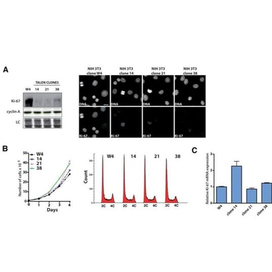
Description: The monoclonal antibody SolA15 recognizes mouse and rat Ki-67, a 300 kDa nuclear protein. Ki-67 is present during all active phases of the cell cycle (G1, S, G2, and mitosis), but is absent from resting cells (G0). Ki-67 is detected within the nucleus during interphase but redistributes to the chromosomes during mitosis. Ki-67 is used as a marker for determining the growth fraction of a given population of cells. In studies of tumor cells, the "Ki-67 labeling index" refers to the number of Ki-67 positive cells within the population and this is used to predict outcome of particular cancer types. Ki-67 has been shown to interact with the DNA-bound protein chromobox protein homolog 3 (CBX3) (heterochromatin).
The SolA15 antibody also recognizes human, non-human primate and canine Ki-67.
Applications Reported: This SolA15 antibody has been reported for use in intracellular staining followed by flow cytometric analysis, immunohistochemical staining of frozen tissue sections, immunohistochemical staining of formalin-fixed paraffin embedded tissue sections, microscopy, and immunocytochemistry.
Applications Tested: This SolA15 antibody has been tested immunocytochemistry of fixed and permeabilized C2C12 cells and can be used at less than or equal to 5 µg/mL or intracellular staining and flow cytometric analysis of stimulated mouse esplenocytes cells using the Foxp3/Transcription Factor Buffer Set (Product # 00-5523-00) and protocol. Please see BestProtocols® Section (Staining intracellular Antigens for Flow Cytometry) for staining protocol (refer to Protocol B: One-step protocol for intracellular (nuclear) proteins). This can be used at less than or equal to 0.125 µg per test. It is recommended that the antibody be carefully titrated for optimal performance in the assay of interest.
Filtration: 0.2 µm post-manufacturing filtered.
靶标信息
Ki-67 is a nuclear protein that is expressed during various stages in the cell cycle, particularly during late G1, S, G2, and M phases. The protein has a forkhead associated domain (FHA) through which it associates with euchromatin at the perichromosomal layer, the centromeric heterochromatin, and the nucleolus. Ki-67 is shown to have a cell cycle dependent topographical distribution with perinucleolar expression at G1, expression in the nuclear matrix at G2, and expression on the chromosomes during M phase. Ki-67 is commonly used as a proliferation marker because it is not detected in G0 cells, but increases steadily from G1 through mitosis. Ki-67 antibodies are useful in establishing the cell growing fraction in neoplasms. In neoplastic tissues, the prognostic value is comparable to the tritiated thymidine-labelling index. The correlation between low Ki-67 index and histologically low-grade tumors is strong. Ki-67 is routinely used as a neuronal marker of cell cycling and proliferation.
应用案例
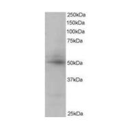
Ki-67 Antibody (13-5698-82) in WB
Ki-67 Antibody (13-5698-82) in ICC/IF
Ki-67 Antibody (13-5698-82) in WB, ICC/IF
风险提示:丁香通仅作为第三方平台,为商家信息发布提供平台空间。用户咨询产品时请注意保护个人信息及财产安全,合理判断,谨慎选购商品,商家和用户对交易行为负责。对于医疗器械类产品,请先查证核实企业经营资质和医疗器械产品注册证情况。
 用户评价
用户评价 暂无用户评价
 文献和实验
文献和实验[1] Xiong YJ, Soomro SH, Huang ZH, Yu PP, Ping J, Fu H. Poly-L-ornithine blocks the inhibitory effects of fibronectin on oligodendrocyte differentiation and promotes myelin repair. Neural Regen Res. 2023 Apr;18(4):832-839. doi: 10.4103/1673-5374.353493. PMID: 36204851; PMCID: PMC9700116.
[2] Sullivan DK, Deutzmann A, Yarbrough J, Krishnan MS, Gouw AM, Bellovin DI, Adam SJ, Liefwalker DF, Dhanasekaran R, Felsher DW. MYC oncogene elicits tumorigenesis associated with embryonic, ribosomal biogenesis, and tissue-lineage dedifferentiation gene expression changes. Oncogene. 2022 Nov;41(45):4960-4970. doi: 10.1038/s41388-022-02458-9. Epub 2022 Oct 7. PMID: 36207533; PMCID: PMC10257951.
[3] Zeng X, Liao H, Wang F. MicroRNA-384 inhibits nasopharyngeal carcinoma growth and metastasis via binding to Smad5 and suppressing the Wnt/β-catenin axis. Cytotechnology. 2021 Apr;73(2):203-215. doi: 10.1007/s10616-021-00458-3. Epub 2021 Feb 26. PMID: 33911345; PMCID: PMC8035371.
of choice (15µg, Lipofectamine 2000™ Invitrogen). The following day the flag-tagged protein expressing plasmid transfected cultures are transfected again with 100nM EF52 biotin labeled siRNA (antisense or sense alone) using Lipofectamine 2000™ or stock 3.4µM
expressing plasmid of choice (15µg, Lipofectamine 2000™ Invitrogen). The following day the flag-tagged protein expressing plasmid transfected cultures are transfected again with 100nM EF52 biotin labeled siRNA (antisense or sense alone) using Lipofectamine
secondary antibody review -- data from 99 publications
cytometry used as a control to detect cell responses targeted antigen 7 Alexa Fluor 488 7 Cy3 8 goat IgG Alexa Fluor 488 1:2000 detect antibody binding in human embryonic kidney 293T cells Invitrogen 9 donkey
 技术资料
技术资料暂无技术资料 索取技术资料

