


- ¥2312
- AAT Bioquest
- 进口
- 22804
- 2026年05月21日

企业认证
相关产品推荐更多 >


万千商家帮你免费找货
0 人在求购买到急需产品
- 详细信息
- 文献和实验
- 技术资料
- 英文名:
Cell Meter™ Mitochondrion Membrane Potential Assay Kit *Orange Fluorescence Optimized for Flow Cytometry*
- 库存:
50
- 供应商:
广州市左克生物科技发展有限公司
- 规格:
100Tests
| Ex (nm) | - | Em (nm) | - |
| 分子量 | - | 溶剂 | - |
| 存储条件 | - |
Cell Meter 线粒体膜电位检测试剂盒是美国AAT Bioquest生产的用于线粒体膜电位检测的试剂盒,Cell Meter检测试剂盒是一类用于检测细胞功能的系列工具,包括细胞活性、细胞毒性、细胞凋亡、细胞膜电位以及细胞周期等方面的指标。每种检测方案均能提供不同荧光颜色的检测方案。这些检测方案为从多角度研究细胞功能活动提供了一种十分有效的方法。Cell Meter线粒体膜电位检测试剂盒 *橙色荧光 适合流式细胞检测* 专门通过检测线粒体跨膜电势的减少来检测细胞凋亡。线粒体跨膜电势瓦解与线粒体渗透转运孔的开放相一致,导致细胞色素C释放到胞浆中,它是下游凋亡级联反应的触发器。该荧光实验方案利用我们独有的MitoLite Orange来检测凋亡细胞中线粒体跨膜电势的减少。在正常细胞中,当MitoLite Orange积聚于线粒体时,红色荧光增加。然而在凋亡细胞中,MitoLite Orange随着线粒体跨膜电位的塌陷而减少。被MitoLite Orange染色的细胞可以通过488n的激发光(FL2 通道)来观察。该试剂盒可以与其它Cell Meter检测试剂盒同时检测细胞的活性和凋亡,该试剂盒已被优化,可用于流式细胞法筛选凋亡抑制剂。,为您提供优质的Cell Meter 线粒体膜电位检测试剂盒。
点击查看光谱
适用仪器
| 流式细胞仪 | |
| Ex: | 488 nm or 532 nm |
| Em: | 575/26 nm |
| 通道: | PE 通道 |
样品实验方案
简要概述
1.用密度为5×10⁵到1×10⁶个细胞/ mL的测试化合物制备细胞
2.将2μL500XMitoTell Orange加入1 mL细胞溶液中
3.将细胞在37℃,5%CO2培养箱中孵育15-30分钟
4.将细胞沉淀,并将细胞重悬于1mL生长培养基中
5.使用具有FL2通道的流式细胞仪分析细胞
操作步骤
1.对于每个样品,将细胞制备在1 mL温热培养基或您选择的缓冲液中,密度为5×10⁵至1×10⁶个细胞/ mL。注意:应对每个细胞系进行单独评估,以确定诱导细胞凋亡的细胞密度。
2.用测试化合物处理细胞一段所需的时间以诱导细胞凋亡,并建立平行对照实验。
3.对于阴性对照:仅用载体处理细胞。
4.对于阳性对照:使用FCCP或CCCP以5-50μM在37℃,5%CO2培养箱中处理细胞15至30分钟。注意:CCCP或FCCP可与MitoTell Orange同时添加。为了获得结果,可能需要对每个单独的细胞系滴定CCCP或FCCP。
5.将2μL500XMitoTell 橙(组分A)加入处理过的细胞中。
6.将细胞在37℃,5%CO2培养箱中孵育15至30分钟。注意:对于粘附细胞,用0.5 mM EDTA轻轻提起细胞以保持细胞完整,并在用MitoTell Orange染料加载溶液孵育之前用含有血清的培养基洗涤细胞一次。
7.将细胞以1000rpm离心4分钟,然后将细胞重悬于1mL的分析缓冲液(组分B)或您选择的缓冲液中。
8.使用具有FL2通道(Ex / Em = 540 / 590nm)的流式细胞仪监测荧光强度。
数据分析
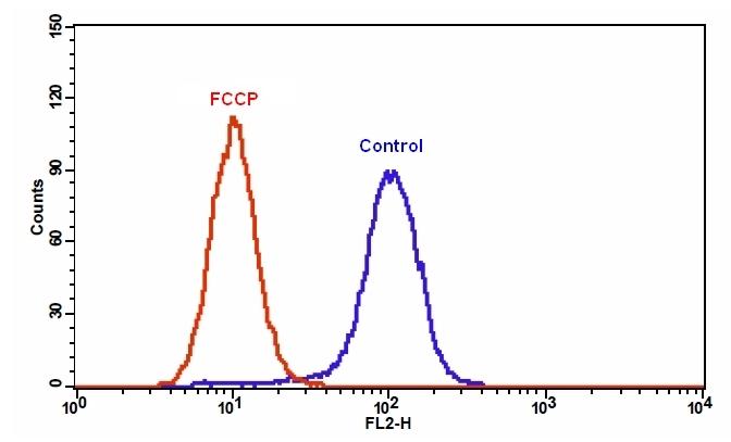
| 图1.在Jurkat细胞中添加FCCP后MitoTell Orange的荧光强度降低。 将Jurkat细胞单独加载MitoTell Orange(蓝色)或在30μMFCCP(红色)存在下加载15分钟。 使用FL2通道,用FACSCalibur(Becton Dickinson,San Jose,CA)流式细胞仪测量MitoTell Orange的荧光强度。 |
参考文献
Safranine O as a fluorescent probe for mitochondrial membrane potential studied on the single particle level and in suspension
Authors: Perevoshchikova IV, Sorochkina AI, Zorov DB, Antonenko YN.
Journal: Biochemistry (Mosc) (2009): 663
Computer-assisted live cell analysis of mitochondrial membrane potential, morphology and calcium handling
Authors: Koopman WJ, Distelmaier F, Esseling JJ, Smeitink JA, Willems PH.
Journal: Methods (2008): 304
Determination of high mitochondrial membrane potential in spermatozoa loaded with the mitochondrial probe 5,5',6,6'-tetrachloro-1,1',3,3'-tetraethylbenzimidazolyl-carbocyanine iodide (JC-1) by using fluorescence-activated flow cytometry
Authors: Guthrie HD, Welch GR.
Journal: Methods Mol Biol (2008): 89
Effects of eprosartan on mitochondrial membrane potential and H2O2 levels in leucocytes in hypertension
Authors: Labios M, Martinez M, Gabriel F, Guiral V, Ruiz-Aja S, Beltran B, Munoz A.
Journal: J Hum Hypertens (2008): 493
How DASPMI reveals mitochondrial membrane potential: fluorescence decay kinetics and steady-state anisotropy in living cells
Authors: Ramadass R, Bereiter-Hahn J.
Journal: Biophys J (2008): 4068
Life cell quantification of mitochondrial membrane potential at the single organelle level
Authors: Distelmaier F, Koopman WJ, Testa ER, de Jong AS, Swarts HG, Mayatepek E, Smeitink JA, Willems PH.
Journal: Cytometry A (2008): 129
Mitochondrial membrane potential in axons increases with local nerve growth factor or semaphorin signaling
Authors: Verburg J, Hollenbeck PJ.
Journal: J Neurosci (2008): 8306
The mitochondrial membrane potential and Ca2+ oscillations in smooth muscle
Authors: Chalmers S, McCarron JG.
Journal: J Cell Sci (2008): 75
[Evaluation of sperm mitochondrial membrane potential by JC-1 fluorescent staining and flow cytometry]
Authors: Xia XY, Wu YM, Hou BS, Yang B, Pan LJ, Shi YC, Jin BF, Shao Y, Cui YX, Huang YF.
Journal: Zhonghua Nan Ke Xue (2008): 135
Cyclosporin A-induced oxidative stress is not the consequence of an increase in mitochondrial membrane potential
Authors: van der Toorn M, Kauffman HF, van der Deen M, Slebos DJ, Koeter GH, Gans RO, Bakker SJ.
Journal: Febs J (2007): 3003
相关产品
| 产品名称 | 货号 |
| Cell Meter 线粒体膜电位检测试剂盒 红色荧光 适合流式细胞检测 | Cat#22806 |
| Cell Meter 线粒体膜电位检测试剂盒 橙色荧光 适合微孔板检测 | Cat#22805 |
| Cell Meter 线粒体膜电位检测试剂盒 红色荧光 适合微孔板检测 | Cat#22807 |
风险提示:丁香通仅作为第三方平台,为商家信息发布提供平台空间。用户咨询产品时请注意保护个人信息及财产安全,合理判断,谨慎选购商品,商家和用户对交易行为负责。对于医疗器械类产品,请先查证核实企业经营资质和医疗器械产品注册证情况。
 文献和实验
文献和实验染料的浓度,引起假去极化。荧光探针JC-1是一种阳离子型的亲脂性染料,能够自由穿过细胞膜,随细胞膜电位的变化而在膜两侧保持动态平衡。其特点是线粒体膜电位低时浓度低,主要以单体形式存在,488nm激发时最大发射波长为527nm,呈绿色荧光,胞浆相对线粒体为低电位,形成流式图中所有细胞FL1均为阳性;膜电位高时浓度高形成聚集体,488nm激发时的最大发射波长为590nm,红色荧光。活细胞线粒体膜电位高,线粒体内JC-1聚集体的浓度高,红色荧光很强,在流式图上表现为FL1和FL2双阳性,而凋亡细胞则大多
,0.6~2.0μm大小的荧光微球适合于这一类的研究,如分析大鼠中性粒细胞、人横纹导管细胞、小鼠腹膜巨噬细胞、人多核白细胞的吞噬功能或不同调理素调理作用对吞噬功能的影响等。 (4)血流分析,10-15 μm大小7种颜色的荧光可供研究组织中局部血流情况,如肿瘤脉管血流速率、视网膜和脉络膜循环、肺泡微管的功能直径定位等。 (5)敏感性诊断试剂,如替代一些已开展应用的微球诊断试验:胶乳凝集试验、微球捕获ELASA、双位点夹心法等,它较传统比色方法更为灵敏;另有新近诞生的流式微球分析
: 检测的时候用什么激发光来观察? 是紫外吗?还有就是晚期调亡和死亡如何来鉴别啊? wangzhua: 应当用蓝色激发吧,我用的蓝色,死亡后应当都是橙色或红色,弥散的,大小与原细胞相当.凋亡的应当有染色体聚集 lhxqxj: AO用蓝光激发,显示绿色,EB用绿光激发,显示红色。 zhangql930611: 我用的是绿激发,凋亡早期的细胞着橙色,晚期的着绿色,但是死亡细胞和晚期凋亡的细胞不能区分,用流式细胞检测仪可以区分 AO/EB染色检测贴壁细胞凋亡 jieyuan_gx: 我们准备用细胞
 技术资料
技术资料暂无技术资料 索取技术资料

