


- ¥2888
- AAT Bioquest
- 进口
- 21800
- 2026年04月06日

企业认证
相关产品推荐更多 >


万千商家帮你免费找货
0 人在求购买到急需产品
- 详细信息
- 文献和实验
- 技术资料
- 英文名:
CytoTrace™ Ultra Green
- 库存:
50
- 供应商:
广州市左克生物科技发展有限公司
- 规格:
1mg
| Ex (nm) | - | Em (nm) | - |
| 分子量 | 822.72 | 溶剂 | DMSO |
| 存储条件 | 在零下15度以下保存, 避免光照 |
CytoTrace Ultra Green新一代荧光示踪探针,非常适合跟踪细胞的运动或位置。该染料可以很好地保留在细胞中,并且追踪活细胞数代。与在相同条件下的CMFDA相比,CytoTrace Ultra Green明显更亮,光稳定性更高并且更耐用。在组织固定后,这种染料产生的信号将非常稳定,使其成为与其他类型的分析方法(如细胞毒性等)结合用于多色应用的理想选择。 CytoTrace Ultra Green的激发光谱和发射光谱与FITC相同,并且与常见的红色荧光染料(例如Texas Red,Cy5,Cy7,iFluor 647和750,AlexaFluor®647和750)完全分离。 CytoTrace Ultra Green可轻松用于各种生物应用的活细胞跟踪,并与流式细胞仪和荧光显微镜兼容。,为您提供优质的CytoTrace Ultra Green。
实验方案
简要概述
- 准备实验细胞
- 溶液配制
- 在室温或37℃下将染料与细胞一起孵育15至30分钟
- 去除染料中的工作溶液
- 使用流式细胞仪分析(Ex / Em = 490/520 nm-FITC滤波器)
溶液配制
储备溶液配制
CytoTrace Ultra Green原液(2-10 mM):添加适量的DMSO充分混合以制成CytoTrace Ultra Green原液(2-10 mM)。注意:储备溶液应立即使用;所有剩余的溶液应等分,并在<-20 o C下冷冻。避免重复冻融循环,并避光。
工作溶液配制
CytoTrace Ultra Green工作溶液:通过用Hanks和20 mM Hepes缓冲液(HHBS)或自备的pH=7的缓冲液稀释DMSO储备液,制备0.5至5 µM的染料工作液,并离心。注意:在某些细胞类型,可能需要较低的浓度才能染色细胞。我们建议在进行实验之前针对每种细胞类型进行测试以获得佳浓度使实验更顺利。
操作步骤
- 用测试化合物处理细胞。
- 离心细胞以获得每管2-10×10 5个细胞。
- 将细胞重悬于500 µL CytoTrace Ultra Green工作溶液中。
- 在室温或37°C下,将细胞与染料溶液孵育15至30分钟,避光。
- 从细胞中除去染料工作溶液;可选:4%甲醛固定细胞。
- 用HHBS或自备的缓冲液洗涤细胞一次。
- 将细胞重悬于500 µL预热的HHBS或培养基中,每管可得到2-10×10 5个细胞。
- 使用流式细胞仪或带有FITC滤波片组的荧光显微镜检测Ex / Em = 490/520 nm处的荧光变化。
图示
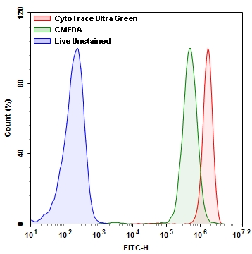
图1. Jurkat细胞中CytoTrace Ultra Green与CMFDA的荧光强度比较。在37 ℃,5%CO2培养箱中,将Jurkat细胞用CytoTrace Ultra Green和CMFDA染料分别加载30分钟。使用带有FITC通道的ACEA NovoCyte 3000流式细胞仪测量荧光强度。
参考文献
Fluorescence-Based Transport Assays Revisited in a Human Renal Proximal Tubule Cell Line
Authors: Caetano-Pinto P, Janssen MJ, Gijzen L, Verscheijden L, Wilmer MJ, Masereeuw R.
Journal: Mol Pharm (2016): 933
The variable chemotherapeutic response of Malabaricone-A in leukemic and solid tumor cell lines depends on the degree of redox imbalance
Authors: Manna A, De Sarkar S, De S, Bauri AK, Chattopadhyay S, Chatterjee M.
Journal: Phytomedicine (2015): 713
Cell membrane tracker based on restriction of intramolecular rotation
Authors: Zhang C, Jin S, Yang K, Xue X, Li Z, Jiang Y, Chen WQ, Dai L, Zou G, Liang XJ.
Journal: ACS Appl Mater Interfaces (2014): 8971
A multiple model probability hypothesis density tracker for time-lapse cell microscopy sequences
Authors: Rezatofighi SH, Gould S, Vo BN, Mele K, Hughes WE, Hartley R.
Journal: Inf Process Med Imaging (2013): 110
Evaluation of stability and sensitivity of cell fluorescent labels when used for cell migration
Authors: Beem E, Segal MS.
Journal: J Fluoresc (2013): 975
TLM-Tracker: software for cell segmentation, tracking and lineage analysis in time-lapse microscopy movies
Authors: Klein J, Leupold S, Biegler I, Biedendieck R, Munch R, Jahn D.
Journal: Bioinformatics (2012): 2276
Horizontal DNA transfer from donor to host cells as an alternative mechanism of epithelial chimerism after allogeneic hematopoietic cell transplantation
Authors: Waterhouse M, Themeli M, Bertz H, Zoumbos N, Finke J, Spyridonidis A.
Journal: Biol Blood Marrow Transplant (2011): 319
The exocytosis of fluorescent nanodiamond and its use as a long-term cell tracker
Authors: Fang CY, Vaijayanthimala V, Cheng CA, Yeh SH, Chang CF, Li CL, Chang HC.
Journal: Small (2011): 3363
The interplay between Leishmania promastigotes and human Natural Killer cells in vitro leads to direct lysis of Leishmania by NK cells and modulation of NK cell activity by Leishmania promastigotes
Authors: Lieke T, Nylen S, Eidsmo L, Schmetz C, Berg L, Akuffo H.
Journal: Parasitology (2011): 1898
Cell electrofusion visualized with fluorescence microscopy
Authors: Trontelj K, Usaj M, Miklavcic D.
Journal: J Vis Exp. (2010)
相关产品
| 货号 # | 产品名称 | 规格 | 分子量 | Ex/Em (nm) | 溶剂 |
| 22014 | CytoTrace Orange CMTMR | 10x50 mg | 554.04 | 541/565 | DMSO |
| 22015 | CytoTrace Red CMPTX | 10x50 mg | 686.25 | 577/602 | DMSO |
| 22016 | CytoTrace Red CFDA | 1 mg | 652.43 | 560/574 | DMSO |
| 22017 | CytoTrace Green CMFDA | 1 mg | 464.86 | 494/521 | DMSO |
| 21800 | CytoTrace Ultra Green | 1 mg | 822.72 | 494/521 | DMSO |
| 22020 | FDA (Fluorescein diacetate) | 1 g | 416.83 | 494/521 | DMSO |
风险提示:丁香通仅作为第三方平台,为商家信息发布提供平台空间。用户咨询产品时请注意保护个人信息及财产安全,合理判断,谨慎选购商品,商家和用户对交易行为负责。对于医疗器械类产品,请先查证核实企业经营资质和医疗器械产品注册证情况。
 文献和实验
文献和实验Green Fluorescent Protein (GFP) as an Indicator of Transfection in Chicken Embryos
Green fluorescent protein (GFP) is responsible for the bioluminescence of the Pacific Northwest jellyfish, Aequorea victoria. In A.victoria, the 27-kDa protein absorbs blue light from a photoprotein that is activated by calcium and emits green
即 Monodansylcadaverine,单丹磺酰尸胺染色,包括自噬体,所有酸性液泡都被染色,故属于非特异性的。 5. CellTrackerTM Green 染色 主要用于双染色,但其能染所有的液泡,故也属于非特异性的。 自噬相关蛋白定位 在研究自噬相关蛋白时,需对其进行定位。由于自噬体与溶酶体、线粒体、内质网、高尔基体关系密切,为了区别,常用到一些示踪蛋白在荧光显微镜下来共定位:Lamp-2:溶酶体膜蛋白,可用于监测自噬体与溶酶体融合。LysoTrackerTM 探针:有红或蓝色可选,显示
毒素+FITC 2.标记细胞器荧光探针 (1)线粒体 Mitochondria Rodamin 123 505/534 ,可染活细胞,阳离子性,可检测线粒体膜电位, 且在多数细胞中停留时间短 JC1 线粒体膜电位低时为单体 490/527 发绿光 线粒体膜电位低时为多聚体 490/590 发红光 可标记活细胞线粒体,且为检测线粒体膜电 位最佳探针: Mitotracker Green FM 490/516, 染活细胞或固定
 技术资料
技术资料暂无技术资料 索取技术资料

