

- 询价
- 美国Advansta公司蛋白质分析试剂
- 美国Advansta公司蛋白质分析试剂
- USA
- 2025年11月23日

企业认证

万千商家帮你免费找货
0 人在求购买到急需产品
- 详细信息
- 询价记录
- 文献和实验
- 技术资料
- 库存:
现货100
- 英文名:
美国Advansta公司蛋白质分析试剂
- 供应商:
上海辅泽商贸有限公司
- 保存条件:
冷藏,-20度
- 规格:
美国Advansta公司蛋白质分析试剂

蛋白质样品加载缓冲液
预混合的标准Laemmli负载缓冲液用于蛋白质电泳
- 预混合- 只需在您的蛋白质样品中添加等量(以2倍提供)
- 获得具有标准化缓冲液的可重现结果
- 从还原或非还原加载缓冲区 中选择
| CAT # | PRODUCT | SIZE | PRICE | |
|---|---|---|---|---|
| R-03018-B10 | Non-reducing protein sample loading buffer (2X) | 1 ml | -/- | |
| R-03018-B50 | Non-reducing protein sample loading buffer (2X) | 5 ml | -/- | |
| R-03147-B10 | Non-reducing protein sample loading buffer (4X) | 1 ml | -/- | |
| R-03147-B50 | Non-reducing protein sample loading buffer (4X) | 5 ml | -/- | |
| R-03148-B10 | Red Non-reducing protein sample loading buffer (4X) | 1 ml | -/- | |
| R-03148-B50 | Red Non-reducing protein sample loading buffer (4X) | 5 ml | -/- | |
| R-03019-B10 | Reducing protein sample loading buffer (2X) | 1 ml | -/- | |
| R-03019-B50 | Reducing protein sample loading buffer (2X) | 5 ml | -/- | |
| R-03146-B10 | Odorless Reducing protein sample loading buffer (2X) | 1 ml | -/- | |
| R-03146-B50 | Odorless Reducing protein sample loading buffer (2X) | 5 ml | -/- |
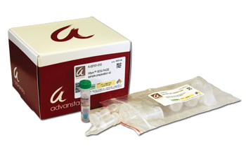
Afyon SDS-PAGE样品制备套件
快速,易于浓度和SDS-PAGE样品纯化的套件
- 纯化- 去除可能干扰电泳(GUHCL,尿素,硫酸铵等)的污染物
- 快速- 准备在不到10分钟内加载的蛋白质样品
- 安全- 无毒 - 无需DMSO
- 与SDS-PAGE和下游Western印迹兼容
- 通过超滤或透析,多次比浓度和缓冲液交换快。
| CAT # | PRODUCT | SIZE | PRICE | |
|---|---|---|---|---|
| K-02101-010 | Afyon SDS-PAGE Sample Preparation Kit | 10 rxns | -/- | |
| K-02101-025 | Afyon SDS-PAGE Sample Preparation Kit | 25 rxns | -/- | |
| R-03020-B10 | Non-reducing protein sample loading buffer (2X) | 1 ml | -/- |
描述
使用Afyon样品纯化套件进行浓缩并纯化蛋白质样品以进行SDS-PAGE
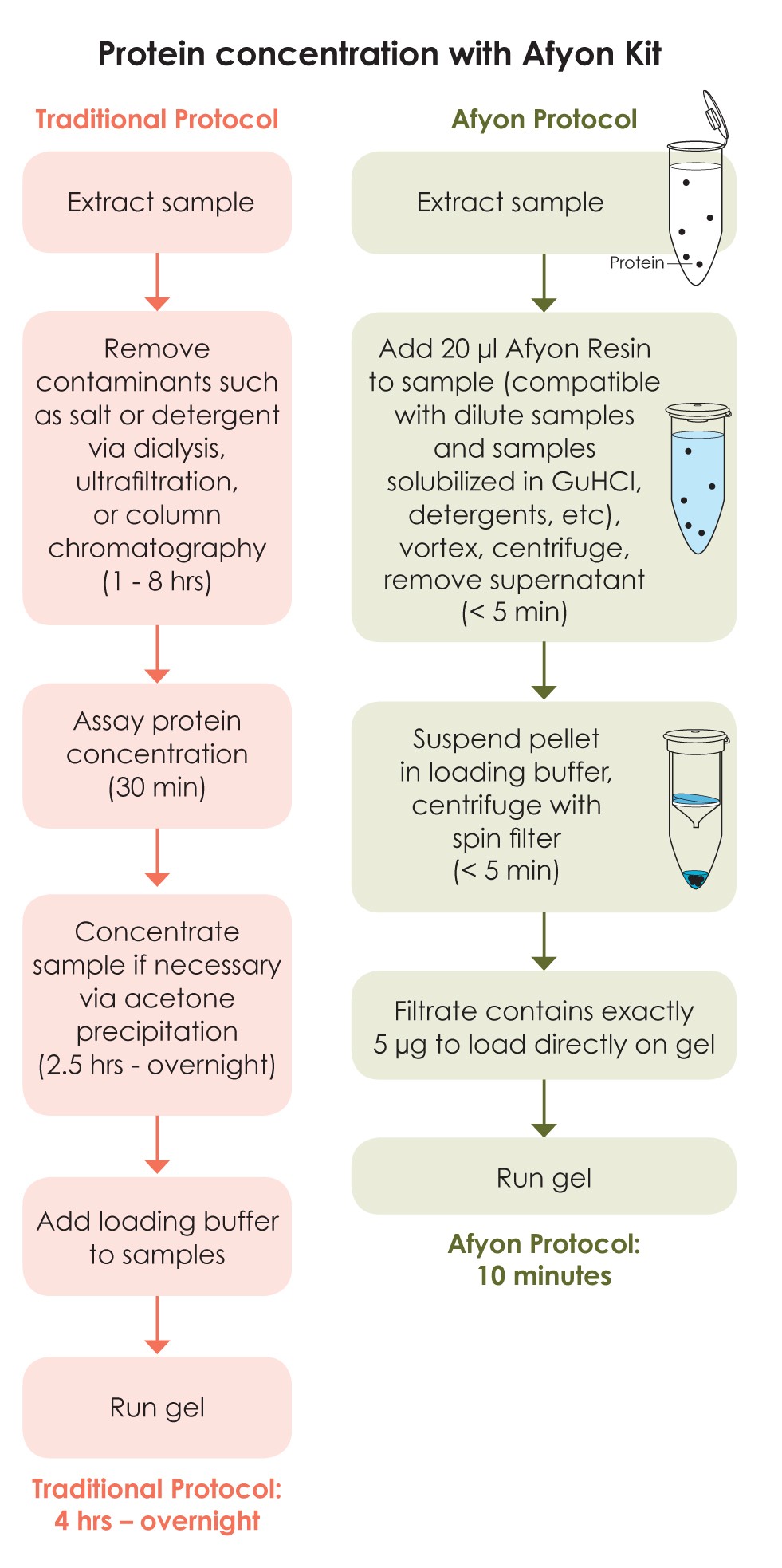
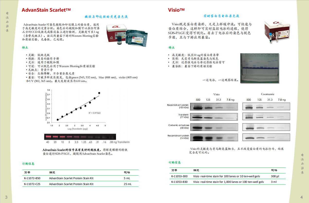
Afyon SDS-PAGE样品制备套件提供了一种快速浓缩蛋白质样品的方法,并将其与干扰电泳的缓冲液分开。快速,有效的协议生成准备在不到十分钟内加载凝胶上的样品(图1),
比使用透析,或TCA沉淀等替代方法可以实现的速度要快得多。此外,AFYON协议很容易扩展,从而使多个样品并行制备。样品与化学发光和荧光蛋白质印迹均保持兼容(图2)。
纯化成功地去除了可以干扰电泳的连体剂(图3)A显示出与超速离心的相似结果,同时十倍(图4)。
在短短10分钟内准备样品。
|
|
图1:与传统纯化协议相比,AFYON协议。 |
Afyon浓度与蛋白质印迹检测兼容。
|
|
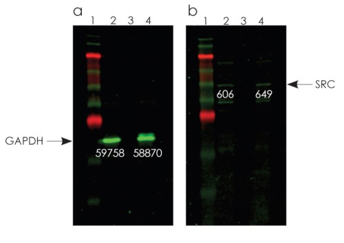
图2:使用A431细胞提取物(面板A和B,泳道2),稀释的细胞提取物(泳道3)的样品创建蛋白质印迹, 并使用Afyon方案(泳道4)浓缩了稀释的提取物。使用Westernbright MCF多色荧光蛋白质印迹套件检测到印迹。 当GAPDH(面板A,泳道3)或SRC(面板B,泳道3)染色时,在稀释的细胞提取物中未检测到条带。但是, 在Afyon浓度之后,对于两种蛋白质,很容易看到带(泳道4)。频段强度表示。
|
Afyon去除可能干扰电泳的污染物。
|
|
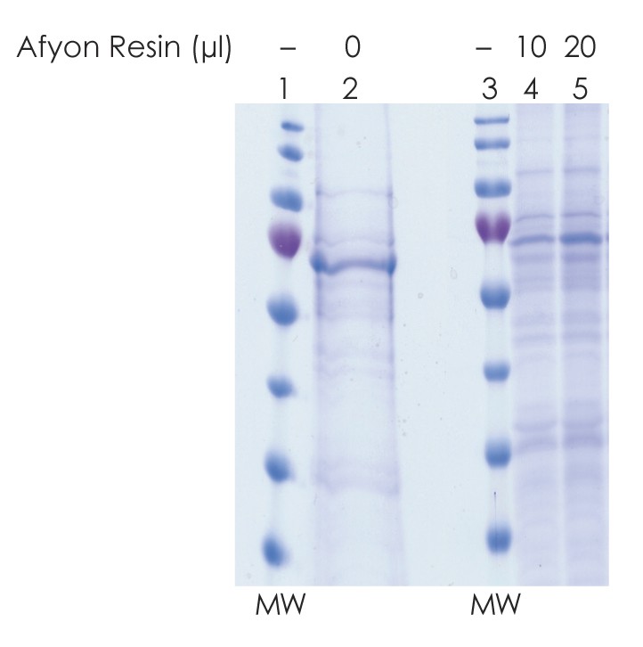
图3: Afyon去除了缓冲液组件,例如氯化鸟苷,硫氰酸酯或尿素,可能导致样品不规则地迁移。 4 m硫丹盐中的K-561细胞裂解物样品散布并在凝胶上不规则地运行(泳道2)。当使用Afyon树脂(小道4-5)回收蛋白质时,将盐除去盐,并干净地运行样品。 10μL的Afyon恢复了2.5μg蛋白(泳道4),而20μL的Afyon恢复了5μg蛋白(泳道5)。 MW =分子量标记。
|
浓缩样品比自旋超滤快10倍。
|
|
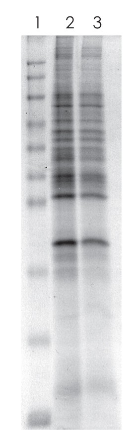
图4:。用1M NaCl,20 mM Tris pH 7.6将HeLa细胞裂解物稀释至20μg/ml。将1 mL稀释的裂解物浓缩,并使用超滤清器自旋滤波器(Milipore)或Afyon树脂交换缓冲液。超滤花了一个多小时,而阿菲恩(Afyon)花了不到10分钟的时间。泳道1:分子量标记。泳道2:通过超滤浓缩的样品。泳道3:使用Afyon浓缩的样品。
|
| # | 产品 | 尺寸 | 价格 | 数量 |
|---|---|---|---|---|
| K-11172-B50 | Advanstain Scarlet蛋白染色套件 | 5ml | - / - | |
| K-111072-C25 | Advanstain Scarlet蛋白染色套件 | 25毫升 | - / - |
描述
Advanstain Scarlet是一种用于凝胶和印迹的荧光蛋白染色,可允许对蛋白质带的敏感和定量可视化。通过快速简便的协议,凝胶可以在不到3个小时内染色,并在各种成像系统上成像。可以在任何荧光成像系统(包括激光和CCD的系统)上成像用Advanstain Scarlet染色的凝胶和印迹,其检测极限小于1 ng的蛋白质或斑点。 Advanstain Scarlet与下游蛋白质印迹或质谱完全兼容,无毒且可生物降解,可安全,简单地处置。
Advanstain Scarlet表现出灵敏度和线性
 |
 |
|
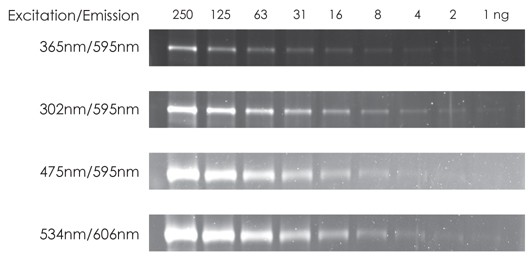
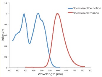
图1。在多个成像条件下可以成像,其灵敏度为1 ng或更少,每频段都可以成像。 Advanstain Scarlet的最佳激发和发射波长与多种成像条件兼容,并且可以使用跨粉刺剂,CCD-或Laser,使用UV(305或365 nm),蓝色或绿光激发使用紫外线(305或365 nm),蓝色或绿光激发,用Advanstain Scarlet染色的凝胶。基于基于成像系统。
|
 |
|
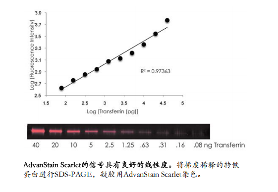
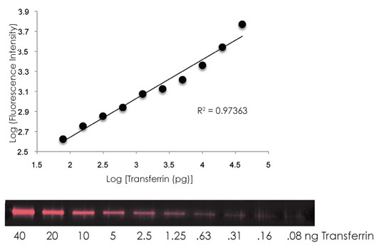
图2。AdvanstainScarlet信号是线性的,对于超过2个数量级。通过SDS-PAGE分析了转铁蛋白蛋白的系列稀释液,并用Advanstain Scarlet染色。
|
Advanstain Scarlet优于其他蛋白质污渍
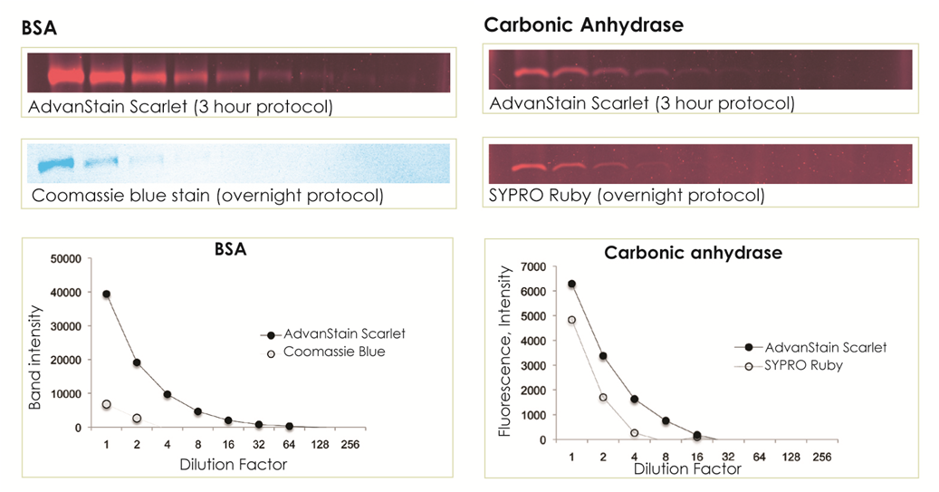
图3。AdvanstainScarlet优于Coomassie和Sypro Ruby。根据制造商的说明,将凝胶用Advanstain Scarlet,Sypro Ruby或Coomassie Blue染色染色,然后在CCD成像器上成像。
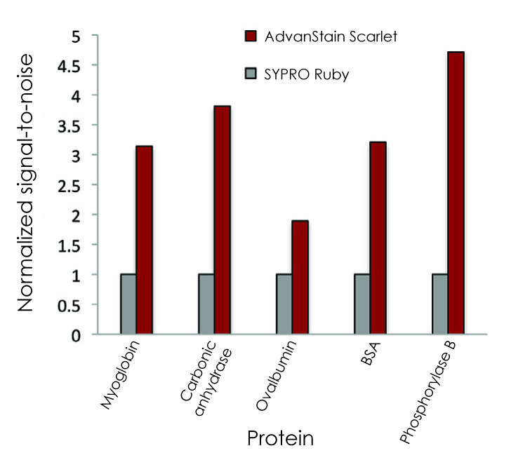
图4。AdvanstainScarlet提供的信噪比比Sypro Ruby更高。 将几种蛋白质的连续稀释液在重复的1D凝胶上分离,
然后根据每个制造商的说明用Advanstain Scarlet或Sypro Ruby染色。对于每种蛋白质,
将信噪比值标准化为Sypro红宝石染色凝胶上蛋白质获得的值。对于每种蛋白质,
Advanstain Scarlet提供了至少比Sypro Ruby的2倍的信噪比。使用AdvanStain观察到的较低背景负责较高的信号到噪声,
从而增加了对低实现波段和斑点定量的信心。
| CAT # | PRODUCT | PRICE | QUANTITY |
|---|---|---|---|
| K-11053-300 | Visio 100 lanes or 10 gels 0.3ml | -/- | |
| K-11053-B30 | Visio 1000 lanes or 100 gels 3ml | -/- |
描述
Visio是一种载荷缓冲液和蛋白质污渍。
Visio在加载前很快与样品中的蛋白质结合,从而导致在凝胶电泳过程中形成可见的带。不需要染色或破坏步骤。 Visio可以实时监测蛋白质电泳,并且与下游质谱完全兼容。
Visio很敏感,方便,不会干扰迁移。
此外,粘膜具有与Coomassie染色相当的高灵敏度(图1),并且不会通过一维凝胶影响蛋白质迁移(图2)。
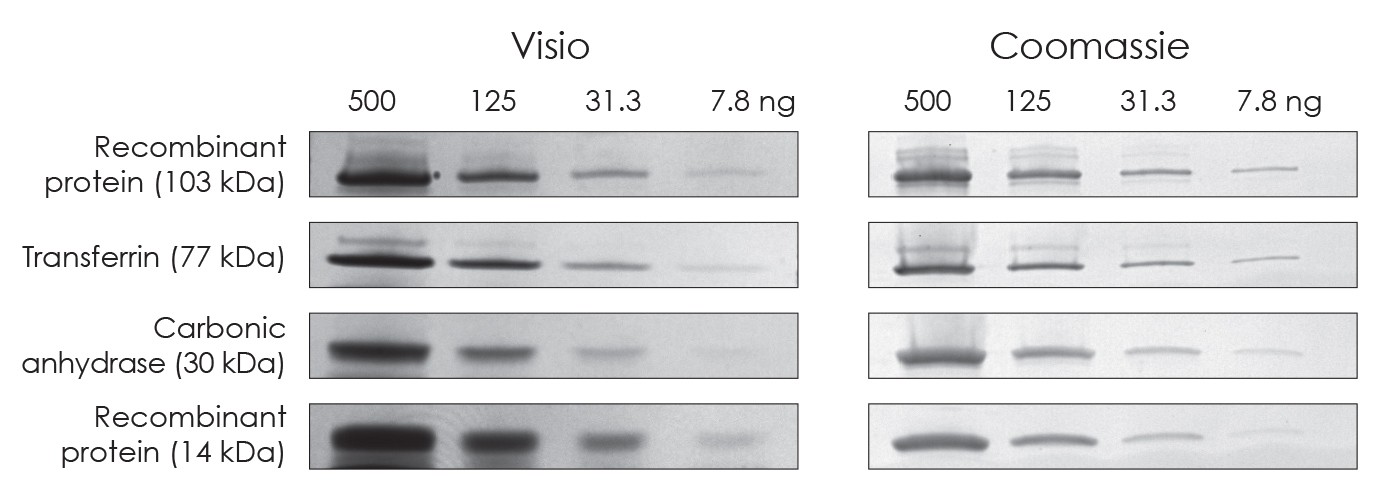
图1:高灵敏度
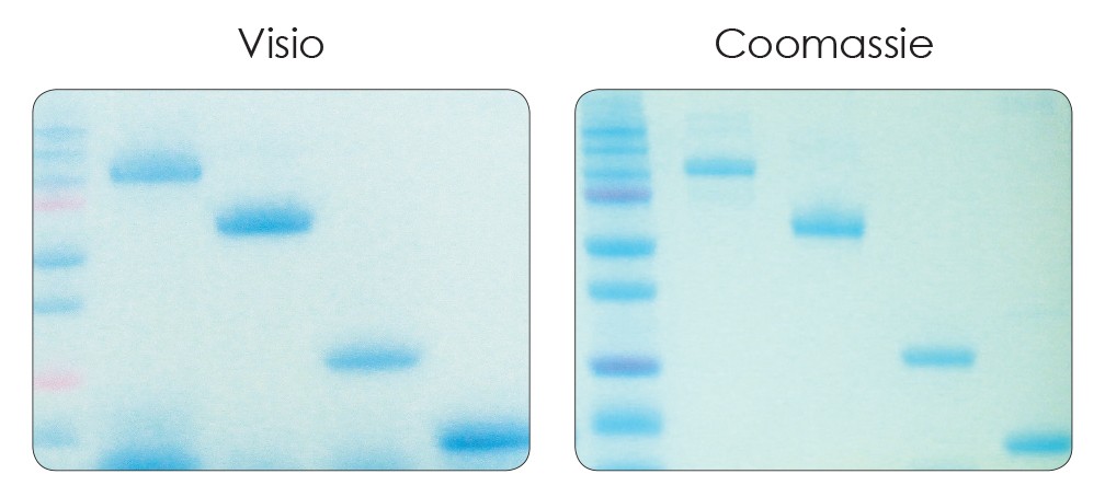
图2:Visio不会影响蛋白质迁移。重复的样品在同一凝胶上运行。左侧的样品在电泳前用粘膜标记,而右侧的凝胶在电泳后用coomassie染色。

| CAT # | PRODUCT | SIZE | PRICE | QUANTITY |
|---|---|---|---|---|
| R-03090-D25 | FlashBlot Transfer Buffer (50x) | 250 ml | -/- | |
| R-03090-D50 | FlashBlot Transfer Buffer (50x) | 500 ml | -/- |
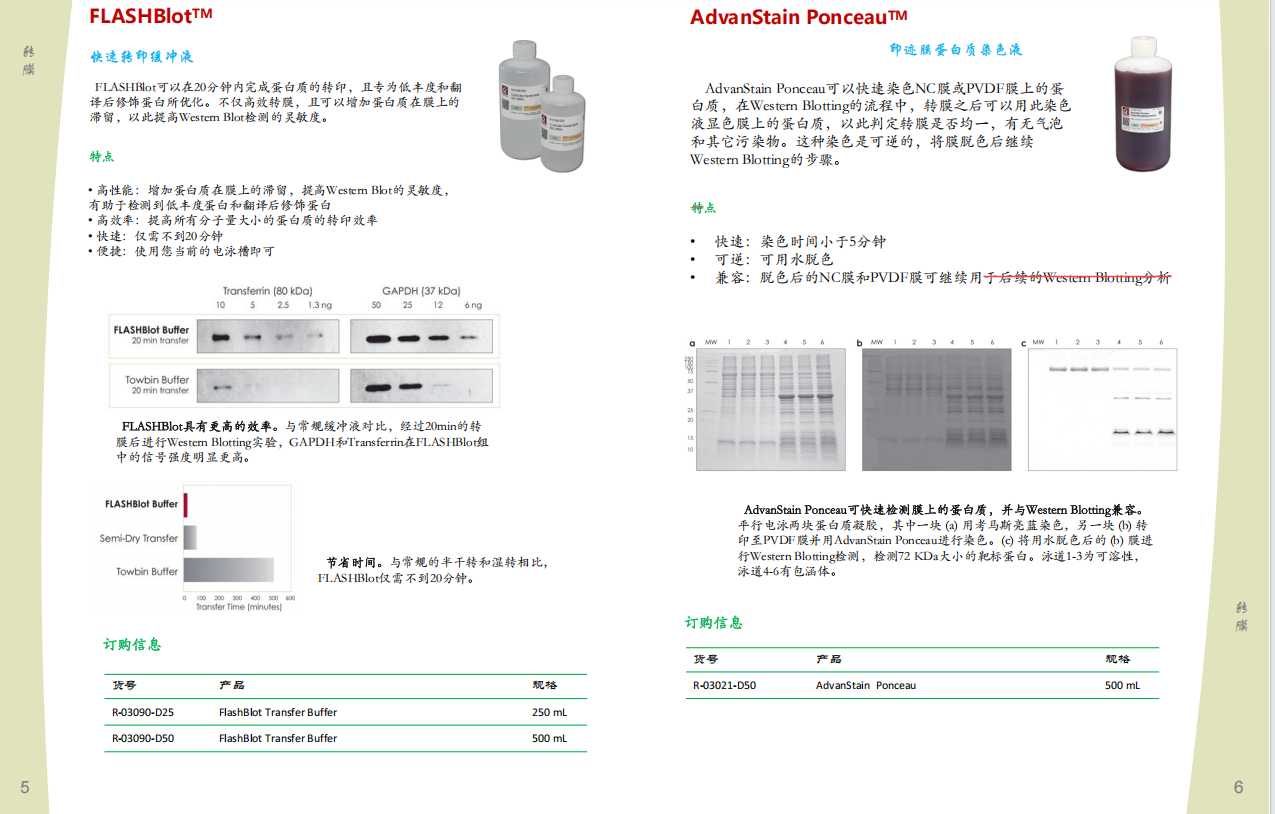
描述
与传统的湿传输缓冲液相比,Flashbrot传递缓冲液提供了所有蛋白质的转移效率,包括高分子重量的蛋白质的转移效率,
这通常很难实现完整的转移。
 |
|
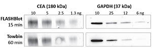
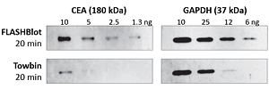
在不到20分钟的时间内完成转移。 使用Flashblot和Towbin传递缓冲液比较了CEA(高MW)和GAPDH(低MW)蛋白的转移。 经过蛋白质印迹分析后,数据显示了15分钟内通过Flashbot与Towbin缓冲液进行的等效蛋白质转移。 |
由于转移时间更快,Flashbrot传递缓冲液可在膜上增加蛋白质的保留,从而更敏感地检测到低含量的蛋白质。
 |
| 较短的传输时间。 Flashbrot使用任何传统的湿传输设备在不到20分钟的时间内实现完整的蛋白质转移。 |
 |
|
通过Flashbrot Buffer更有效地传输。 转移20分钟后,Western印迹分析表明, 使用FlashBlot缓冲液比使用Towbin缓冲液更有效地将CEA和GAPDH更有效地转换。 |
短的转移时间 - 加上转移效率和蛋白质保留率的提高 - 可以对翻译后修饰的蛋白质进行敏感检测,
在传统转移方案后可能难以检测。
 |
|
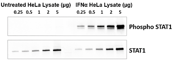
精美的转换后修饰的感兴趣的蛋白质。使用STAT1和Phosho-STAT1对未处理和IFNα处理过的 HELA细胞裂解液的蛋白质印迹分析显示了FlashBlot传递缓冲液的效率。印迹是用CCD相机成像的。 |

| # | 产品 | 尺寸 | 价格 | 数量 |
|---|---|---|---|---|
| R-03090-D25 | Flashbrot传输缓冲区(50x) | 250毫升 | - / - | |
| R-03090-D50 | Flashbrot传输缓冲区(50x) | 500毫升 | - / - |
描述
与传统的湿传输缓冲液相比,Flashbrot传递缓冲液提供了所有蛋白质的转移效率,包括高分子重量的蛋白质的转移效率,这通常很难实现完整的转移。
 |
| 在不到20分钟的时间内完成转移。 使用Flashblot和Towbin传递缓冲液比较了CEA(高MW)和GAPDH(低MW)蛋白的转移。经过蛋白质印迹分析后,数据显示了15分钟内通过Flashbot与Towbin缓冲液进行的等效蛋白质转移。 |
由于转移时间更快,Flashbrot传递缓冲液可在膜上增加蛋白质的保留,从而更敏感地检测到低含量的蛋白质。
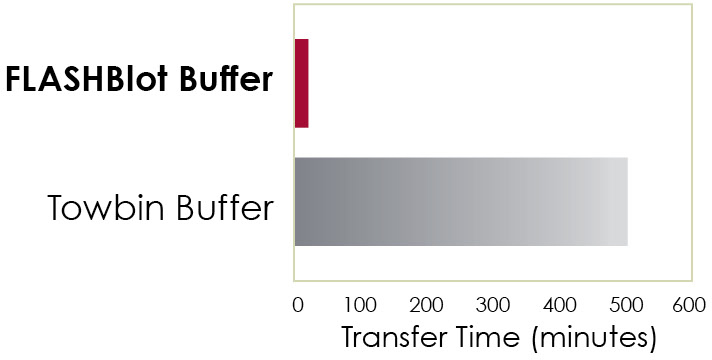 |
| 较短的传输时间。 Flashbrot使用任何传统的湿传输设备在不到20分钟的时间内实现完整的蛋白质转移。 |
 |
|
通过Flashbrot Buffer更有效地传输。 转移20分钟后,Western印迹分析表明, 使用FlashBlot缓冲液比使用Towbin缓冲液更有效地将CEA和GAPDH更有效地转换。 |
短的转移时间 - 加上转移效率和蛋白质保留率的提高 - 可以对翻译后修饰的蛋白质进行敏感检测,在传统转移方案后可能难以检测。
 |
|
精美的转换后修饰的感兴趣的蛋白质。使用STAT1和Phosho-STAT1对未处理和IFNα处理过的 HELA细胞裂解液的蛋白质印迹分析显示了FlashBlot传递缓冲液的效率。印迹是用CCD相机成像的。 |
描述

Westernbright ECL HRP基板
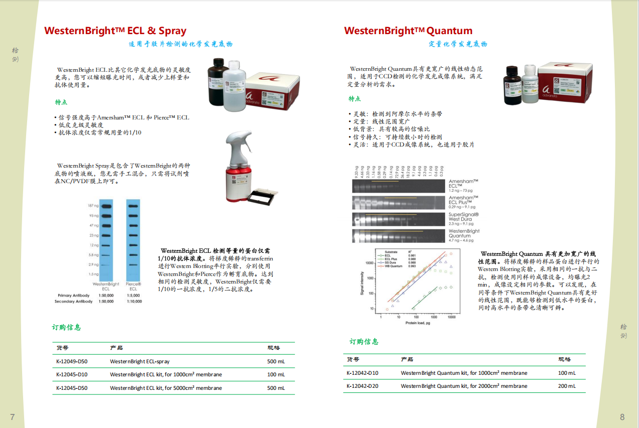
WesternBright®ECL是一种使用X射线膜成像化学发光蛋白质印迹的辣根过氧化物酶底物。 Westernbright ECL比其他化学发光底物更敏感,
并且使用抗体的抗体最多比其他底物少10倍,从而可以节省珍贵的抗体和样品。此外,Westernbright ECL信号持续持久,允许多次暴露而没有实质性信号衰减。
Westernbright ECL针对使用X射线膜成像的化学发光蛋白质印迹进行了优化。
|
|
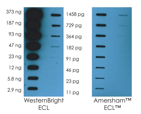
Westernbright ECL专门配制,可为敏感的膜成像产生强信号。 Westernbright ECL底物在与HRP反应后产生强,持久的信号。使用Westernbright ECL或Amersham™ECL™(GE Healthcare)检测到含有转铁蛋白蛋白连续稀释液的重复插槽印迹。两种印迹同时暴露于同一薄膜片15秒钟。 Westernbright ECL比Amersham ECL更敏感。
|
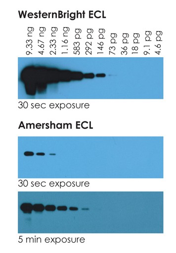
Westernbright ECL对检测低丰度蛋白具有高灵敏度
|
|
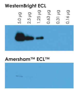
当检测低丰度蛋白质时,西方ECL的较高灵敏度尤为重要。探测了ERK1的双重蛋白质印迹,并使用Westernbright ECL或Amersham ECL HRP底物检测到HeLa细胞裂解物的系列稀释液。每个印迹都暴露于胶卷1分钟。
|
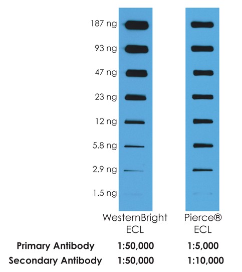
Westernbright ECL所需的抗体最多比其他底物少十倍
|
|
用所示的抗体含量探测了含有转铁蛋白连续稀释液的重复插槽印迹,并根据制造商的说明用Westernbright ECL或Pierce ECL(Thermo Scientific)检测。印迹是在同一部电影上同时成像的。 Westernbright ECL产生的敏感性相同,初级抗体少了十倍,次生抗体却降低了五倍。
|
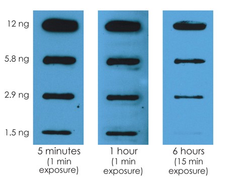
Westernbright ECL信号是长期的,可以长时间暴露或多次暴露,而没有实质性的信号衰减。
|
|
Westernbright ECL产生持久信号。底物孵育六个小时后,可以对Westernbright ECL检测到的印迹。无需急于去暗室来形象印迹。
| |
|
|
Westernbright ECL的更大稳定性允许在长时间暴露期间积累更多的信号,从而提高了检测到非常低的丰度蛋白的能力。对ATP1A1蛋白进行了探测,重复的蛋白质印迹被探测,并使用Westernbright ECL或Amersham ECL检测到。在10分钟的曝光中,只能通过任何一个底物检测到2个频段。在接触60分钟的情况下,在Westernbright ECL印迹的1.25μg车道中可见条带,而信号衰减几乎没有差异 | |
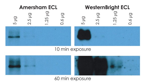
Westernbright ECL的强信号需要较短的较短频带
|
|
Westernbright ECL产生强信号,导致更高的灵敏度,暴露时间较短。根据制造商的说明,使用Westernbright ECL或Amersham ECL检测到含有转铁蛋白蛋白的重复印迹,并将印迹暴露于同一薄膜中。与Amersham ECL印迹相比,Westernbright ECL印迹的30秒暴露能够检测到更多的频带。
|
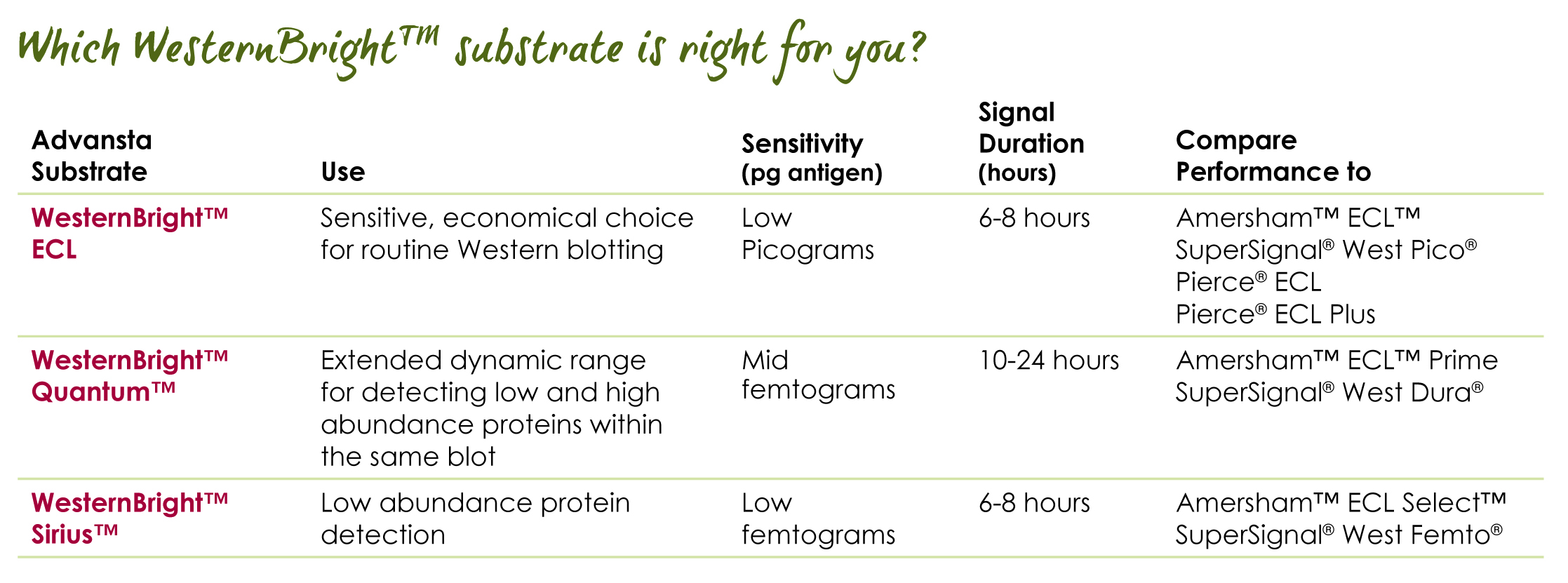
WesternBright Quantum HRP substrate
Quantitative chemiluminescent Western blotting
- Sensitive – detect attomoles of protein per band.
- Quantitative – linear range of signal with respect to protein amount exceeds 3 orders of magnitude
- Versatile – Optimized for CCD imaging, and compatible with film detection. Chemifluorescent emissions can be detected with fluorescence imaging systems
- Low background – for high signal to noise ratio
- Long lasting signal – image blots hours after substrate incubation

| CAT # | PRODUCT | SIZE | PRICE | QUANTITY |
|---|---|---|---|---|
| K-12042-C20 | WesternBright Quantum trial size kit | 20ml | -/- | |
| K-12042-D10 | WesternBright Quantum kit | 100ml | -/- | |
| K-12042-D20 | WesternBright Quantum kit | 200ml | -/- |
Description

WesternBright Quantum HRP substrate
WesternBright® Quantum® chemiluminescent substrate sets the bar for sensitivity and quantitative ability. Specially developed for CCD imaging, WesternBright Quantum produces a strong, long-lasting signal with extremely low background, perfect for detecting low abundance proteins. Since it does not exhibit substrate depletion at high protein loads, WesternBright Quantum provides the largest dynamic range of any chemiluminescent substrate for the most quantitative chemiluminescent Western experiments.
WesternBright Quantum exhibits high sensitivity with the greatest linear range
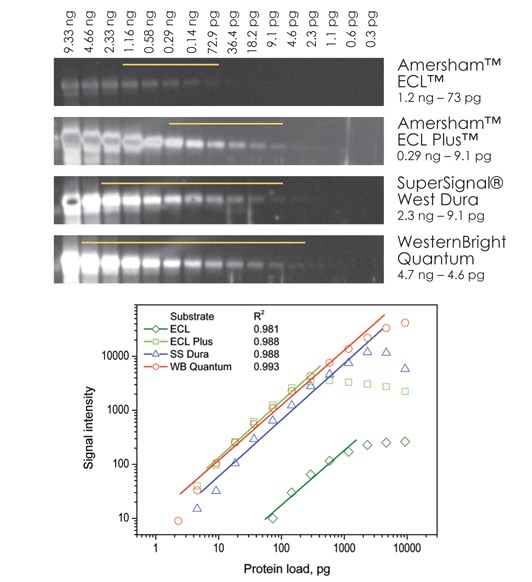
|
|
Identical Western blots containing serial dilutions of transferrin were probed with a rabbit-anti-transferrin primary antibody and a goat-anti-rabbit secondary antibody conjugated to HRP. The blots were incubated with chemiluminescent substrates as recommended by each manufacturer. All blots were imaged simultaneously for 2 min on a CCD imager and display parameters were identical across all images. Band intensities were plotted and a best fit linear regression conducted for each substrate. WesternBright Quantum shows the largest linear dynamic range out of all four substrates with the highest R2 value. Bands that fall on the linear part of the curve for each substrate are indicated in the image. |

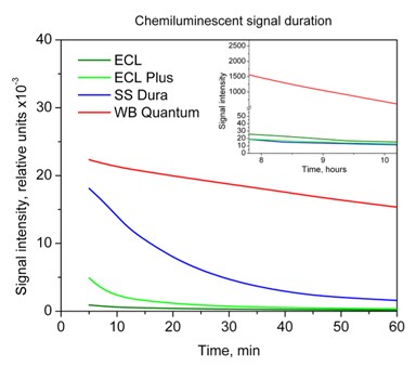
WesternBright Quantum produces the most stable chemiluminescent signal
|
|
Blots detected using WesternBright Quantum or one of three other chemiluminescent substrates were re-imaged at several times over a 10 hr period. The intensitiy of one band is plotted. 60 min after substrate incubation, WesternBright Quantum retains 70% of its initial signal strength, while the competition decays to 5% or less. |

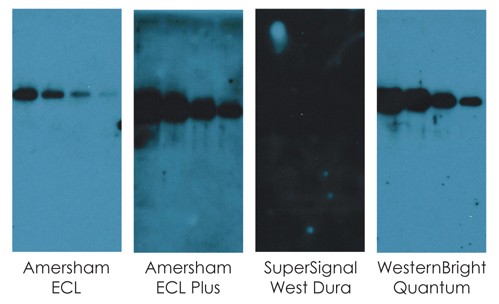
Extremely low background when using WesternBright Quantum
|
|
Replicate Western blots were developed using WesternBright Quantum or one of three other chemiluminescent substrates. After a simultaneous 20 min exposure to the same piece of film, WesternBright Quantum displays the best combination of sensitivity and low background signal. All display parameters were identical across all images shown in this figure. |

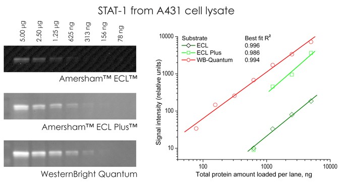
High sensitivity for quantitative detection of low-abundance proteins
|
|
WesternBright Quantum provides the highest sensitivity and broadest linear dynamic range. Serial dilutions of A431 cell lysates were blotted and probed to detect STAT-1 protein. The blots were detected with WesternBright Quantum,or two other chemiluminescent substrates (ECL and ECL Plus, GE Healthcare). Only data points on the linear portion of the best-fit curve for each substrate shown. All display parameters were identical across all images in this figure. |

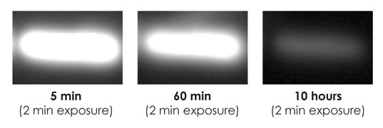
No need to rush to image your Western blot
|
|
WesternBright Quantum allows blots to be imaged several hours after substrate incubation. Blots can be re-imaged to obtain the perfect exposure, without worrying about losing signal. A blot was imaged with 2 min exposures at 5 min, 60 min, and 10 hours after substrate incubation. The same band is clearly seen in each image, even after 10 hours. |
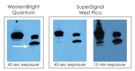
Superior sensitivity with film
|
|
WesternBright Quantum provides high-performance film detection.WesternBright Quantum or SuperSignal® West Pico (Thermo Scientific) were used to detect duplicate Western blots. A band (arrow) is detected by WesternBright Quantum in a brief exposure, while a much longer exposure is needed to detect the same band with SuperSignal West Pico. All display parameters were identical across all images in this figure
|

WesternBright Quantum HRP substrate
Quantitative chemiluminescent Western blotting
- Sensitive – detect attomoles of protein per band.
- Quantitative – linear range of signal with respect to protein amount exceeds 3 orders of magnitude
- Versatile – Optimized for CCD imaging, and compatible with film detection. Chemifluorescent emissions can be detected with fluorescence imaging systems
- Low background – for high signal to noise ratio
- Long lasting signal – image blots hours after substrate incubation
Includes:
WesternBright Quantum luminol/enhancer solution
WesternBright Quantum stabilized peroxide solution
| CAT # | PRODUCT | SIZE | PRICE | QUANTITY |
|---|---|---|---|---|
| K-12042-C20 | WesternBright Quantum trial size kit | 20ml | -/- | |
| K-12042-D10 | WesternBright Quantum kit | 100ml | -/- | |
| K-12042-D20 | WesternBright Quantum kit | 200ml | -/- |
Description
WesternBright Quantum HRP substrate
WesternBright® Quantum® chemiluminescent substrate sets the bar for sensitivity and quantitative ability. Specially developed for CCD imaging, WesternBright Quantum produces a strong, long-lasting signal with extremely low background, perfect for detecting low abundance proteins. Since it does not exhibit substrate depletion at high protein loads, WesternBright Quantum provides the largest dynamic range of any chemiluminescent substrate for the most quantitative chemiluminescent Western experiments.
WesternBright Quantum exhibits high sensitivity with the greatest linear range
|
|
Identical Western blots containing serial dilutions of transferrin were probed with a rabbit-anti-transferrin primary antibody and a goat-anti-rabbit secondary antibody conjugated to HRP. The blots were incubated with chemiluminescent substrates as recommended by each manufacturer. All blots were imaged simultaneously for 2 min on a CCD imager and display parameters were identical across all images. Band intensities were plotted and a best fit linear regression conducted for each substrate. WesternBright Quantum shows the largest linear dynamic range out of all four substrates with the highest R2 value. Bands that fall on the linear part of the curve for each substrate are indicated in the image. |
WesternBright Quantum produces the most stable chemiluminescent signal
|
|
Blots detected using WesternBright Quantum or one of three other chemiluminescent substrates were re-imaged at several times over a 10 hr period. The intensitiy of one band is plotted. 60 min after substrate incubation, WesternBright Quantum retains 70% of its initial signal strength, while the competition decays to 5% or less. |
Extremely low background when using WesternBright Quantum
|
|
Replicate Western blots were developed using WesternBright Quantum or one of three other chemiluminescent substrates. After a simultaneous 20 min exposure to the same piece of film, WesternBright Quantum displays the best combination of sensitivity and low background signal. All display parameters were identical across all images shown in this figure. |
High sensitivity for quantitative detection of low-abundance proteins
|
|
WesternBright Quantum provides the highest sensitivity and broadest linear dynamic range. Serial dilutions of A431 cell lysates were blotted and probed to detect STAT-1 protein. The blots were detected with WesternBright Quantum,or two other chemiluminescent substrates (ECL and ECL Plus, GE Healthcare). Only data points on the linear portion of the best-fit curve for each substrate shown. All display parameters were identical across all images in this figure. |
No need to rush to image your Western blot
|
|
WesternBright Quantum allows blots to be imaged several hours after substrate incubation. Blots can be re-imaged to obtain the perfect exposure, without worrying about losing signal. A blot was imaged with 2 min exposures at 5 min, 60 min, and 10 hours after substrate incubation. The same band is clearly seen in each image, even after 10 hours. |

Superior sensitivity with film
|
|
WesternBright Quantum provides high-performance film detection.WesternBright Quantum or SuperSignal® West Pico (Thermo Scientific) were used to detect duplicate Western blots. A band (arrow) is detected by WesternBright Quantum in a brief exposure, while a much longer exposure is needed to detect the same band with SuperSignal West Pico. All display parameters were identical across all images in this figure
|
WesternBright MCF
Multi-color fluorescent Western blotting in one kit
Try multicolor fluorescent western blotting today. Our kit includes everything except primary antibodies.
- Multiplexing – visualize two proteins simultaneously!
- Sensitivity –10x greater sensitivity than Cy dyes; pictogram detection
- Fast results – 3.5 hours for entire protocol
- Compatible with imaging systems that detect Cy3 and Cy5 (MCF) or near-IR imaging systems (MCF-IR)
- Fluorescent Detection – choose between visible (APC and RPE) or near IR (IR700 and IR800) kits
Secondary antibodies
Blocking solution
Washing solution
Pre-cut low-autofluorescence PVDF membranes
Background quenching sheets
| CAT # | PRODUCT | SIZE | PRICE | QUANTITY |
|---|---|---|---|---|
| K-12023-010 | WesternBright MCF-IR fluorescent Western blotting kit, Goat-anti-mouse IgG IR700/ Goat-anti-rabbit IgG IR800 | 10 assays | -/- | |
| K-12022-010 | WesternBright MCF-IR fluorescent Western blotting kit, Goat-anti-rabbit IgG IR700/ Goat-anti-mouse IgG IR800 | 10 assays | -/- |
Description
WesternBright MCF visible and near infrared (IR) fluorescent Western blotting kits allow the assay of two proteins at once, increasing the quality
and quantity of information that can be gained from a single blot.
Two colors, two proteins, simultaneously
By using dyes that have different spectral properties (excitation and emission), you can visualize them in different channels at the same time on a digital imager.

Figure 1. Antibodies conjugated with different dyes allow you to visualize two antigens at once. A. The diagram represents the basics of a western blot
assay using multi-color fluorescence. b-e. The absorption (gray lines) and emission spectra of the dyes available in the WesternBright kit.
Also known as multiplexing, simultaneous detection with different antibodies is ideal for certain types of experiments. For example, assay a
loading control alongside a protein of interest; assay for two proteins you want to quantify relative to each other; or, assay phosphorylated and non-phosphorylated isoforms of a protein simultaneously.

Figure 2. Simultaneous detection of EGFR and phospho-EGFR with WesternBright MCF. Increased phosphorylation of EGFR
in response to EGF was detected. Lysates from A431 cells (lane 1) and A431 cells treated with EGF (lane 2) were blotted and EGFR
detected in the green channel (b) or phospho-EGFR detected in the red channel (c). The two channels are superimposed in (a). Lane 3:
molecular weight markers.
WesternBright MCF is more sensitive than other Cy dyes
The fluorescent dyes provided with WesternBright MCF outperform Cy dyes and allow detection of low picogram amounts of protein.
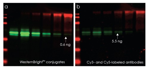
Figure 3. WesternBright Conjugates provide a brighter signal than the ECL Plex Western Blotting system. Duplicate
Western blots containing samples of AFP and CEA proteins were treated identically and probed with the same mixture of mouse
anti-AFP and rabbit anti-CEA primary antibodies. One blot was stained with WesternBright conjugates and the other with an
identical concentration of Cy3 anti-mouse and Cy5 anti-rabbit antibodies, following the protocol recommended for ECL Plex.
Under identical imaging conditions, WesternBright MCF provides a brighter signal, and 10x greater sensitivity.
Additionally, the WesternBright protocol saves time and money since there is no need to strip and re-probe a blot, no use of
disposable film, and the blot can be imaged immediately, without drying. Use the WesternBright multicolor fluorescent Western
blotting kit to try fluorescent Western blotting today.
WesternBright MCF
Multi-color fluorescent Western blotting in one kit
Try multicolor fluorescent western blotting today. Our kit includes everything except primary antibodies.
- Multiplexing – visualize two proteins simultaneously!
- Sensitivity –10x greater sensitivity than Cy dyes; pictogram detection
- Quality Reagents – includes blocking and for washing
- Fast results – 3.5 hours for entire protocol
- Compatible with imaging systems that detect Cy3 and Cy5 (MCF) or near-IR imaging systems (MCF-IR)
- Fluorescent Detection – choose between visible (APC and RPE) or near IR (IR700 and IR800) kits with
Kit Includes:
Secondary antibodies
Blocking solution
Washing solution
Pre-cut low-autofluorescence PVDF membranes
Background quenching sheets
| CAT # | PRODUCT | SIZE | PRICE | QUANTITY |
|---|---|---|---|---|
| K-12023-010 | WesternBright MCF-IR fluorescent Western blotting kit, Goat-anti-mouse IgG IR700/ Goat-anti-rabbit IgG IR800 | 10 assays | -/- | |
| K-12022-010 | WesternBright MCF-IR fluorescent Western blotting kit, Goat-anti-rabbit IgG IR700/ Goat-anti-mouse IgG IR800 | 10 assays | -/- |
Description
WesternBright MCF visible and near infrared (IR) fluorescent Western blotting kits allow the assay of two proteins at once, increasing the quality and quantity of information that can be gained from a single blot.
Two colors, two proteins, simultaneously
By using dyes that have different spectral properties (excitation and emission), you can visualize them in different channels at the same time on a digital imager.
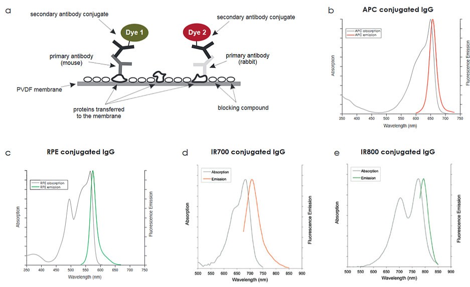
Figure 1. Antibodies conjugated with different dyes allow you to visualize two antigens at once. A. The diagram represents the basics of a western blot assay using
multi-color fluorescence. b-e. The absorption (gray lines) and emission spectra of the dyes available in the WesternBright kit.
Also known as multiplexing, simultaneous detection with different antibodies is ideal for certain types of experiments. For example, assay a loading control alongside a protein
of interest; assay for two proteins you want to quantify relative to each other; or, assay phosphorylated and non-phosphorylated isoforms of a protein simultaneously.
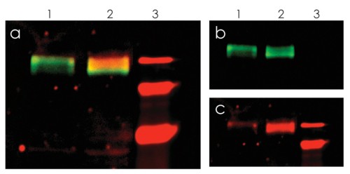
Figure 2. Simultaneous detection of EGFR and phospho-EGFR with WesternBright MCF. Increased phosphorylation of EGFR in response to EGF was detected.
Lysates from A431 cells (lane 1) and A431 cells treated with EGF (lane 2) were blotted and EGFR detected in the green channel (b) or phospho-EGFR detected in the
red channel (c). The two channels are superimposed in (a). Lane 3: molecular weight markers.
WesternBright MCF is more sensitive than other Cy dyes
The fluorescent dyes provided with WesternBright MCF outperform Cy dyes and allow detection of low picogram amounts of protein.

Figure 3. WesternBright Conjugates provide a brighter signal than the ECL Plex Western Blotting system. Duplicate Western blots containing samples of
AFP and CEA proteins were treated identically and probed with the same mixture of mouse anti-AFP and rabbit anti-CEA primary antibodies. One blot was stained
with WesternBright conjugates and the other with an identical concentration of Cy3 anti-mouse and Cy5 anti-rabbit antibodies, following the protocol recommended
for ECL Plex. Under identical imaging conditions, WesternBright MCF provides a brighter signal, and 10x greater sensitivity.
Additionally, the WesternBright protocol saves time and money since there is no need to strip and re-probe a blot, no use of disposable film, and the blot can be
imaged immediately, without drying. Use the WesternBright multicolor fluorescent Western blotting kit to try fluorescent Western blotting today.
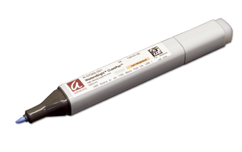
WesternBright ChemiPen
A chemiluminescent marker for writing on membranes
- Improve accuracy - transform your visible protein ladder standard into a chemiluminescent standard on your Western blot
- Annotate - permanently mark your blots with the date or blot ID
- Compatible - with both X-ray film and CCD imagers
- Confirm HRP substrate stability - the ChemiPen reagent glows when incubated with an HRP substrate, so it can be used to test substrate stability
| CAT # | PRODUCT | SIZE | PRICE | QUANTITY |
|---|---|---|---|---|
| R-07055-001 | WesternBright ChemiPen | 1 pen | -/- |
Description
Write or draw on your transfer membranes with the WesternBright® ChemiPen. The reagent in the ChemiPen adsorbs to nitrocellulose and PVDF membranes, and reacts with HRP substrates to produce chemiluminescence that can be detected with X-ray film or CCD imaging.
With the proprietary "ink" you can transform your visible protein markers into chemiluminescent markers, to annotate your blot with a date or blot ID, or to check the stability of your HRP substrate (Figure 1).
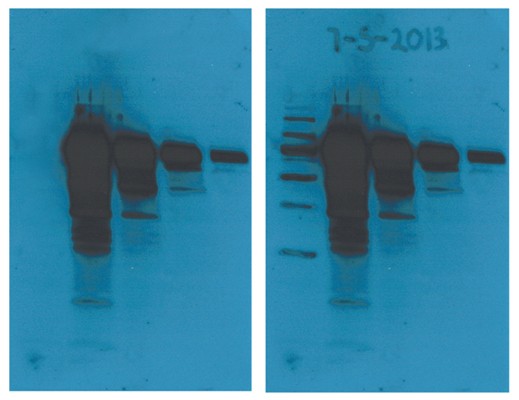
Figure 1. The WesternBright ChemiPen can be used to mark the location of protein standards and to annotate the blot with a date, as in the right panel.
The WesternBright ChemiPen has two tips, a fine tip at one end that is perfect for writing on the blot or tracing the bands of a visible protein standard, and a thicker wedge tip that can be used to deposit greater amounts of reagent on the blot. Marking the location of visible protein standards on a blot is easy. Simply trace the visible markers on the membrane after transfer and before blocking (Figure 2).
ELISABright Chemiluminescent substrate
Chemiluminescent substrate for ELISA applications
- Highest signal to noise ratio – for superior performance
- Broad linear dynamic range – for enhanced detection and precision
- Immediate light generation – for fast detection
- Cost effective – and reduced reagent consumption
Includes:
ELISABright Luminol reagent
ELISABright Peroxide reagent
| CAT # | PRODUCT | SIZE | PRICE | QUANTITY |
|---|---|---|---|---|
| K-16025-D10 | ELISABright substrate | 100 mL | -/- | |
| K-16025-D25 | ELISABright substrate | 250 mL | -/- |
Description
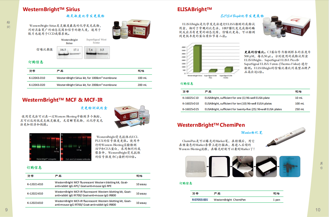
ELISABright is an enhanced and highly sensitive chemiluminescent HRP substrate optimized for chemiluminescent ELISA applications.
This luminol-based substrate demonstrates a wide linear dynamic range (Figure 1) and a superior signal to noise ratio (Figure 2) for
excellent quantitative ability. ELISABright allows for enhanced detection of low abundance proteins and reduced consumption of antibodies and other reagents.
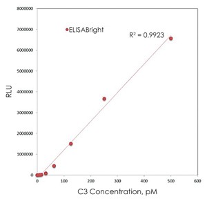 |
Figure 1. ELISABright has a broad linear dynamic range. C3 protein was detected using a sandwich-style ELISA. Plates were coated with anti-C3 antibody and blocked with 1% BSA-PBST. 50 microliters of a series of C3 solutions, ranging in concentration from 7.8 to 500 pM, was added to each well, and C3 protein was detected with a monoclonal anti-huC3a antibody followed by a goat anti-mouse HRP conjugate. Detection was performed according to the ELISABright protocol, using 100 microliters of substrate per well. |
|
Figure 2. ELISABright has a superior signal-to-noise ratio. ELISABright, SuperSignal® ELISA Pico and SuperSignal ELISA Femto substrates (Thermo Fisher) were used to develop replicate sandwich-style ELISA assays as described in Figure 1. Signal-to-noise ratios for detection of wells containing 50 microliters of a 500 pM solution of C3 protein are shown. ELISABright demonstrates a signal-to-noise ratio 6-times higher than the competition. | 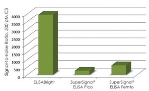 |
风险提示:丁香通仅作为第三方平台,为商家信息发布提供平台空间。用户咨询产品时请注意保护个人信息及财产安全,合理判断,谨慎选购商品,商家和用户对交易行为负责。对于医疗器械类产品,请先查证核实企业经营资质和医疗器械产品注册证情况。
- 作者
- 内容
- 询问日期
 文献和实验
文献和实验生产,如Motorola、HP、IBM、Hitachi等公司。而蛋白质芯片由于在基础研究和技术实现方面尚有诸多难题,因此其商业化步子还很小,据称美国加州Ciphergen生物系统公司已有产品投放市场,而英国的Cambridge Antibody Technology也在利用蛋白质芯片从事研究、诊断和药物发现工作。研究中,我们对国际上知名的43家生物芯片公司的主要从业领域进行了统计(注:有的公司经营多种业务),发现从事分析软件的实际与开发的公司是数量最多的,其次是扫描仪的生产与制造,两者的总和占生物芯片总业务的占30
仪给基因组学带来革命性的影响,4700蛋白质组分析仪必将给后基因组学的研究带来同样的革命性影响。4700 TOF/TOF系统现已成为蛋白组研究的最前沿核心工具。 ICAT蛋白差异表达分析试剂 随着蛋白质组学研究的日益兴起,蛋白质的定性分析技术如测序、 PMF 测定等和仪器如蛋白质测序仪和 MALDI-TOF 、 LC/MS/MS 等已基本成熟,但是人们更关注蛋白质的定量表达分析技术和对低丰度调控蛋白的鉴定。美国应用生物系统公司最新推出了使用 MALDI-TOF 和 LC
化物质检测等。 美国著名细胞产品公司Cell BioLabs为您提供完备的细胞氧化应激分析解决方案,其产品包含蛋白质羰基化、硝基化损伤及晚期氧化蛋白产物(AOPP)分析,脂质过氧化的标志物壬烯(HNE)和丙二醛(MDA)以及8-异前列腺素F2a快速检测试剂盒,核酸DNA/RNA的常规损伤分析如8-OHdG/8-OHG和AP位点检测,还包括细胞水平的彗星分析和DNA双链断裂分析试剂盒;除此之外,还有多种抗氧化物的活性检测方案供您选择,如过氧化物歧化酶(SOD)、过氧化氢酶 (CAT)及ORAC指标


