

- ¥850 - 2150
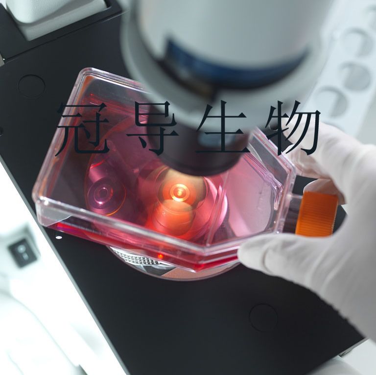
- 冠导生物
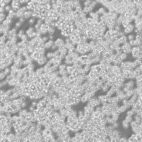
- T24[T-24]人膀胱移行细胞癌传代细胞活性强|送STR图谱
- 美国、德国、欧洲等地
- 2025年07月14日

企业认证
相关产品推荐更多 >


万千商家帮你免费找货
0 人在求购买到急需产品
- 详细信息
- 文献和实验
- 技术资料
- 品系:
详见细胞说明资料
- 细胞类型:
详见细胞说明资料
- 肿瘤类型:
详见细胞说明资料
- 供应商:
上海冠导生物工程有限公司
- 库存:
≥100瓶
- 生长状态:
详见细胞说明资料
- 年限:
详见细胞说明资料
- 运输方式:
常温运输【复苏细胞】或干冰运输【冻存细胞】
- 器官来源:
详见细胞说明资料
- 是否是肿瘤细胞:
详见细胞说明资料
- 细胞形态:
详见细胞说明资料
- 免疫类型:
详见细胞说明资料
- 物种来源:
详见细胞说明资料
- 相关疾病:
详见细胞说明资料
- 组织来源:
详见细胞说明资料
- 英文名:
T24[T-24]人膀胱移行细胞癌传代细胞活性强|送STR图谱
- 规格:
1*10(6)Cellls/瓶
传代方法:1:2-1:4(首次传代建议1:2)
生长特性:贴壁生长
换液频率:每周2-3次
背景资料:该细胞源自一位81岁白人女性患者的膀胱移行细胞癌组织;来源于移行细胞癌病人的白血病和血浆对T24和相关细胞株有细胞毒性;倍增时间为19小时;含ras(H-ras)癌基因,表达肿瘤特有抗原。
U343 Cells;背景说明:详见相关文献介绍;传代方法:1:2传代;生长特性:贴壁生长 ;形态特性:详见产品说明;相关产品有:HEK AD293 Cells、FO [Mouse myeloma] Cells、SKNBE(2) Cells
SNU-520 Cells;背景说明:详见相关文献介绍;传代方法:1:2-1:3传代;每周换液2-3次。;生长特性:贴壁或悬浮,详见产品说明部分;形态特性:详见产品说明;相关产品有:H-1648 Cells、H-460 Cells、TE-3A Cells
HuH28 Cells;背景说明:肝内胆管癌;传代方法:1:2-1:3传代;每周换液2-3次。;生长特性:贴壁;形态特性:详见产品说明;相关产品有:IHH4 Cells、LS 513 Cells、PTK-2 Cells
解冻细胞出现大量细胞碎片的可能原因及推荐解决方案如下:1)冷冻时细胞密度过低:推荐的解决方案:缩短解冻过程中的时间。将细胞取出后,立即将冻存管浸入在37℃的水浴中,并震荡至全部融化,然后迅速地转移到预热的培养基中;2)不适当的冷冻过程:推荐的解决方案:解冻不同冷冻室总的试管。确保在冻存过程中采用的适当的技术,并确保使用了适量和适合的冷冻。出现生长缓慢的可能原因及推荐解决方案如下:1)培养瓶的大小:一般解决方案是一些细胞在培养基中倾向于维持一定的密度。将培养物转移到较小的培养瓶中,使细胞密度上升;如,根据培养基的体积,从75培养瓶转移到2或3个25培养瓶中;2)细胞密度过低:通常解决方案是提GAO未来冷冻物种细胞的冻存密度,或使用较小的培养容器,或解冻多个试管来进行培养。
T24[T-24]人膀胱移行细胞癌传代细胞活性强|送STR图谱
┈订┈购┈热┈线:1┈5┈8┈0┈0┈5┈7┈6┈8┈6┈7【微信同号】┈Q┈Q:3┈3┈0┈7┈2┈0┈4┈2┈7┈1;
产品包装形式:复苏细胞:T25培养瓶(一瓶)或冻存细胞:1ml冻存管(两支)
来源说明:细胞主要来源ATCC、DSMZ等细胞库
物种来源:Human\Mouse\Rat\Others
Panc 04.03 Cells;背景说明:详见相关文献介绍;传代方法:1:2传代;生长特性:贴壁生长;形态特性:上皮样;相关产品有:Mono-Mac-1 Cells、T2(174 x CEM.T2) Cells、L428 Cells
D283-MED Cells;背景说明:详见相关文献介绍;传代方法:每周换液2-3次。;生长特性:悬浮细胞的多细胞聚集体,和一些贴壁 Cells;形态特性:上皮细胞;相关产品有:MBVP Cells、CAL 78 Cells、WM115-mel Cells
IGR-OV1 Cells;背景说明:详见相关文献介绍;传代方法:1:2-1:3传代;每周换液2-3次。;生长特性:贴壁或悬浮,详见产品说明部分;形态特性:详见产品说明;相关产品有:H1734 Cells、EMT6 Cells、MFD-1 Cells
ETCC-007 Cells;背景说明:原位导管癌;女性;传代方法:1:2-1:3传代;每周换液2-3次。;生长特性:贴壁;形态特性:详见产品说明;相关产品有:HEL-92-1-7 Cells、UT-7 Cells、HCC1187 Cells
购买的细胞死亡或细胞存活率不佳可能原因?研究人员在细胞培养时出现存活率不佳,原因比较复杂,常见原因可归纳为:培养基使用错误或培养基品质不佳;血清使用错误或血清的品质不佳;解冻过程错误;冷冻细胞解冻后,加以洗涤细胞和离心;悬浑细胞误认为死细胞;培养温度使用错误;细胞置于-80℃太久等。建议严格参照AC的标准操作规程进行细胞复苏、冻存等工作。欲将一般动物细胞离心下来,其离心速率应为多少转速?欲回收动物细胞,其离心速率一般为300×g(约1OOOrpm),5-10分钟,转速过GAO或离心时间过长都将造成细胞死亡。合适的离心转速是根据相对离心力决定。RCF=1.119×10⁵×r×(rpm)2,其中r为离心机转轴中心与离心套管底部内壁的距离;rpm为离心机每分钟的转数;RCF(relaive nrifugal for)为相对离心力,以重力加速度g(980.66cm/s2)的倍数来表示单位。
┈订┈购┈热┈线:1┈5┈8┈0┈0┈5┈7┈6┈8┈6┈7【微信同号】┈Q┈Q:3┈3┈0┈7┈2┈0┈4┈2┈7┈1;
T24[T-24]人膀胱移行细胞癌传代细胞活性强|送STR图谱
形态特性:上皮细胞样
在细胞培养操作中,每一个步骤都可能影响细胞系的命运。有时,细胞换液后突然死亡,这让科研人员困惑不已。那么,究竟是什么原因导致了这种情况呢?首先,换液操作过程中的不当处理是一个常见因素。例如,使用的移液器如果没有校准准确,吸取或添加培养液的量过多或过少,都可能使细胞所处的环境渗透压发生变化。细胞在渗透压失衡的环境中,水分子会快速进出细胞,导致细胞肿胀或皱缩,最终死亡。此外,如果在吸取旧培养液时过于靠近细胞层,容易造成细胞的机械性损伤,破坏细胞的完整性,使其无法维持正常的生理功能。其次,培养液的成分和质量也至关重要。新配制的培养液若在成分比例上出现偏差,如某些营养物质浓度过高或过低,可能无法满足细胞的生长需求,导致细胞因营养缺乏或中毒而死亡。而且,培养液若在储存或处理过程中受到污染,从而迅速致使细胞死亡。再者,培养环境的变化不容忽视。换液时,比如,温度过高会使细胞内蛋白质变性,温度过低则会降低细胞的活性和代谢速率;二氧化碳浓度的改变会影响培养液的酸碱度,进而干扰细胞的正常生理活动。所以细胞换液后死亡是多种因素综合作用的结果。
HuCCT-1 Cells;背景说明:详见相关文献介绍;传代方法:1:4传代;生长特性:贴壁生长;形态特性:上皮细胞样;相关产品有:T-98 Cells、AR-42J Cells、RPMI1788 Cells
T173 Cells;背景说明:详见相关文献介绍;传代方法:1:2-1:4传代;每周换液2-3次。;生长特性:贴壁生长;形态特性:成纤维细胞;相关产品有:Hep-G2/C3A Cells、GOS3 Cells、SUNE-1 Cells
X63-Ag8.653 Cells;背景说明:详见相关文献介绍;传代方法:1:2-1:3传代;每周换液2-3次。;生长特性:贴壁或悬浮,详见产品说明部分;形态特性:详见产品说明;相关产品有:LU451 Cells、H-196 Cells、SHIN3 Cells
HuT 102 Cells;背景说明:详见相关文献介绍;传代方法:1:3传代,2-3天传一代;生长特性:悬浮生长 ;形态特性:圆形;淋巴母细胞样;相关产品有:CAKI 1 Cells、GM15452 Cells、SW954 Cells
UMR 106 Cells;背景说明:注射放射性同位素磷(32P)诱导产生的可移植性大鼠骨肉瘤克隆建立了UMR-106细胞株。 细胞对PTH,前列腺素及破骨甾体有响应。 对PTH的响应度,UMR-106比相关细胞株UMR-108(ATCC CRL-1663)要高。 蛋白激酶C的活化抑制ATP诱导的胞内水平的升高。 起始骨肉瘤和克隆株都是University of Sheffield的T.J. Martin建立的。;传代方法:1:2-1:3传代;每周换液2-3次。;生长特性:贴壁;形态特性:上皮细胞样;相关产品有:SW 1990 Cells、BSC1 Cells、PC-3M-IE8 Cells
MPP-89 Cells;背景说明:详见相关文献介绍;传代方法:1:2-1:3传代;每周换液2-3次。;生长特性:贴壁或悬浮,详见产品说明部分;形态特性:详见产品说明;相关产品有:Adeno-293 Cells、COLO 738 Cells、IBRS2 Cells
KYSE 510 Cells;背景说明:食管鳞癌;女性;传代方法:1:2-1:3传代;每周换液2-3次。;生长特性:贴壁;形态特性:详见产品说明;相关产品有:NCIH1105 Cells、RK 13 Cells、Hs737T Cells
Hs27F Cells;背景说明:包皮;成纤维细胞;男性;传代方法:1:2-1:3传代;每周换液2-3次。;生长特性:贴壁;形态特性:详见产品说明;相关产品有:LWnt3A Cells、IPLB-Sf21-AE Cells、HLEpiC Cells
Hep2 Cells;背景说明:最初认为这个细胞源自喉上皮癌,但随后通过同功酶分析、HeLa标记染色体和DNA指纹分析发现,起源细胞已被HeLa污染。 角蛋白免疫过氧化物酶染色阳性。;传代方法:1:2传代;生长特性:贴壁生长;形态特性:上皮细胞样;相关产品有:H-660 Cells、hFOB1.19 Cells、LICR-LON-HN6-R Cells
11P0-1 Cells(拥有STR基因鉴定图谱)
Abcam HeLa BAX KO Cells(拥有STR基因鉴定图谱)
AG14859 Cells(拥有STR基因鉴定图谱)
BayGenomics ES cell line RRE061 Cells(拥有STR基因鉴定图谱)
BayGenomics ES cell line XF586 Cells(拥有STR基因鉴定图谱)
┈订┈购┈热┈线:1┈5┈8┈0┈0┈5┈7┈6┈8┈6┈7【微信同号】┈Q┈Q:3┈3┈0┈7┈2┈0┈4┈2┈7┈1;
BY00846 Cells(拥有STR基因鉴定图谱)
CU-19 Cells(拥有STR基因鉴定图谱)
DM319 Cells(拥有STR基因鉴定图谱)
GM03184 Cells(拥有STR基因鉴定图谱)
PANC403 Cells;背景说明:详见相关文献介绍;传代方法:1:2传代;生长特性:贴壁生长;形态特性:上皮样;相关产品有:MFM223 Cells、Hs294T Cells、H-929 Cells
H-2291 Cells;背景说明:详见相关文献介绍;传代方法:1:3-1:4传代;每周换液2-3次。;生长特性:贴壁生长;形态特性:上皮细胞样;相关产品有:SuDHL 4 Cells、BCaP-37 Cells、HeLa/DDP Cells
LWnt-3A Cells;背景说明:详见相关文献介绍;传代方法:1:2-1:3传代;每周换液2-3次。;生长特性:贴壁或悬浮,详见产品说明部分;形态特性:详见产品说明;相关产品有:A549 ATCC Cells、Epstein-Barr-3 Cells、DMS 114 Cells
SNU-1040 Cells;背景说明:结肠癌;男性;传代方法:1:2-1:3传代;每周换液2-3次。;生长特性:贴壁;形态特性:详见产品说明;相关产品有:SCL-2 Cells、MSC Cells、SW-1353 Cells
COLO 320 Cells;背景说明:详见相关文献介绍;传代方法:1:2-1:3传代;每周换液2-3次。;生长特性:贴壁;形态特性:淋巴母细胞;相关产品有:THLE-2 Cells、UM-UC-3 Cells、NCI-H1876 Cells
118MG Cells;背景说明:注意: 据报道来自不同个体的胶质母细胞瘤细胞株U-118 MG (HTB-15) 和 U-138 MG (HTB-16)有着一致的VNTR和相近的STR模式。 U-118 MG 和 U-138 MG细胞遗传学上很相似并有至少六个衍生标记染色体。 这是1966年至1969年间J. Ponten和同事从恶性神经胶质瘤中构建的细胞株中的一株(其它包括ATCC HTB-14和 ATCC HTB-16 and ATCC HTB-17)。 1987年用BM-Cycline培养6周去除了支原体污染。 ;传代方法: 消化3-5分钟。1:2传代。3天内可长满。;生长特性:贴壁生长;形态特性:混合型;相关产品有:SUP-B1 Cells、Detroit 562 Cells、LM1 Cells
Bovine ENDometrial cells Cells;背景说明:子宫内膜;上皮细胞;雌性;传代方法:1:2-1:3传代;每周换液2-3次。;生长特性:贴壁;形态特性:详见产品说明;相关产品有:N9 Cells、MDA-MB-134-VI Cells、CF Pac1 Cells
Hs-600-T Cells;背景说明:详见相关文献介绍;传代方法:1:2-1:3传代,2-3天换液1次。;生长特性:贴壁生长;形态特性:详见产品说明;相关产品有:H-1781 Cells、TMK1 Cells、FM88 Cells
OVCAR 5 Cells;背景说明:卵巢癌;腹水转移;女性;传代方法:1:2-1:3传代;每周换液2-3次。;生长特性:贴壁;形态特性:详见产品说明;相关产品有:Nb-2 Cells、MTC-TT Cells、SW527 Cells
NCI-H2108 Cells;背景说明:详见相关文献介绍;传代方法:1:2-1:3传代;每周换液2-3次。;生长特性:贴壁或悬浮,详见产品说明部分;形态特性:详见产品说明;相关产品有:COLO 684 Cells、D6P2T Cells、University of Arizona Cell Culture-893 Cells
LNT-229 Cells;背景说明:详见相关文献介绍;传代方法:1:4-1:6传代;每周换液2-3次;生长特性:贴壁生长;形态特性:上皮细胞;相关产品有:H1435 Cells、MAVER-1 Cells、C-33A Cells
Eca-109 Cells;背景说明:1973年建系,来自人食管中段鳞癌组织,小块法原代培养建系。BALB/c裸鼠移植成瘤。;传代方法:1:2-1:3传代;每周换液2-3次。;生长特性:贴壁;形态特性:上皮细胞样;相关产品有:SKMEL1 Cells、F-36P Cells、HCMEC(BL12-H) Cells
BC-020 Cells;背景说明:详见相关文献介绍;传代方法:1:2-1:3传代;每周换液2-3次。;生长特性:贴壁或悬浮,详见产品说明部分;形态特性:详见产品说明;相关产品有:C-8161 Cells、Vero E6 Cells、B-16 Cells
5C8C4 Cells(拥有STR基因鉴定图谱)
KE 37 Cells;背景说明:急性T淋巴细胞白血病;男性;传代方法:1:2-1:3传代;每周换液2-3次。;生长特性:悬浮;形态特性:详见产品说明;相关产品有:SU-DHL-4 Cells、NCI-H747 Cells、KP-N-RT Cells
T24[T-24]人膀胱移行细胞癌传代细胞活性强|送STR图谱
HP615 Cells;背景说明:肺癌;传代方法:1:2-1:3传代;每周换液2-3次。;生长特性:贴壁;形态特性:详见产品说明;相关产品有:Panc 8.13 Cells、HPASMC Cells、PIG1 Cells
IA-LM Cells;背景说明:详见相关文献介绍;传代方法:1:2-1:3传代;每周换液2-3次。;生长特性:贴壁或悬浮,详见产品说明部分;形态特性:详见产品说明;相关产品有:CCD-19Lu Cells、H2009 Cells、H-220 Cells
LX-2 Cells;背景说明:详见相关文献介绍;传代方法:1:3传代,2-3天换液一次;生长特性:贴壁生长;形态特性:详见产品说明;相关产品有:MDA-MB-435S Cells、OVCA433_Bast Cells、GM04678 Cells
D407 RPE Cells;背景说明:详见相关文献介绍;传代方法:1:2传代;生长特性:贴壁生长 ;形态特性:详见产品说明;相关产品有:NCI H508 Cells、SuDHL 10 Cells、GM05384 Cells
COLO-320HSR Cells;背景说明:该细胞1984年建系,源自一位33岁患有大肠腺癌男性经5-fu治疗后的腹水。;传代方法:1:2传代。3天内可长满。;生长特性:半贴壁生长;形态特性:详见产品说明;相关产品有:UACC-812 Cells、G-401 Cells、NIH3T3 Cells
DU 145 Cells;背景说明:DU 145 是从一位有3年淋巴细胞白血病史的前列腺癌患者的脑部转移灶中建立的。该细胞系未检测到激素敏感性,酸性酶阳性,单个的细胞可在软琼脂中形成集落。对此细胞和原始肿瘤的亚显微结构分析可见微绒毛、微丝、细胞桥粒、线粒体、发达的高尔基体和异质溶酶体。该细胞不表达前列腺抗原。;传代方法:1:2-1:3传代;每周换液2-3次。;生长特性:贴壁;形态特性:上皮细胞样;相关产品有:Fetal Human Lens-124 Cells、McA-RH 8994 Cells、U118-MG Cells
SNU-354 Cells;背景说明:详见相关文献介绍;传代方法:1:2传代;生长特性:贴壁生长;形态特性:上皮细胞样;相关产品有:SK RC 20 Cells、Ketr-3 Cells、NCTC-1469 Cells
GM15877 Cells(拥有STR基因鉴定图谱)
HAP1 GPD2 (-) 3 Cells(拥有STR基因鉴定图谱)
Kit 225-K6 Cells;背景说明:详见相关文献介绍;传代方法:1:2-1:3传代;每周换液2-3次。;生长特性:贴壁或悬浮,详见产品说明部分;形态特性:详见产品说明;相关产品有:3T3-F442A Cells、PIEC Cells、SK-MEL-3 Cells
Pt K2 (NBL-5) Cells;背景说明:详见相关文献介绍;传代方法:1:2-1:3传代;每周换液2-3次。;生长特性:贴壁或悬浮,详见产品说明部分;形态特性:详见产品说明;相关产品有:NP69SV40T Cells、Cor L88 Cells、NCIH1650 Cells
NCI-H-69 Cells;背景说明:详见相关文献介绍;传代方法:1:2—1:4传代,每周换液2次;生长特性:悬浮生长,聚团;形态特性:聚团悬浮;相关产品有:DHL-8 Cells、HCT116 Cells、Fao Cells
MHCC 97-L Cells;背景说明:来源于中山医院,生长较缓慢;传代方法:1:2传代;生长特性:贴壁生长;形态特性:上皮样;相关产品有:CT-26 WT Cells、U-2OS Cells、Colon38 Cells
H1623 Cells;背景说明:详见相关文献介绍;传代方法:每周换液2-3次。;生长特性:贴壁生长;形态特性:详见产品说明;相关产品有:OCILY7 Cells、HO-8910PM Cells、mouse Inner Medullary Collecting Duct-3 Cells
SK-N-BE Cells;背景说明:1972年11月从一们多次化疗及放疗的扩散性神经母细胞瘤患儿骨髓穿刺物中建立了SK-N-BE(2)神经母细胞瘤细胞株。 该细胞显示中等水平的多巴胺-β-羟基酶活性。 有报道称SK-N-BE(2)细胞的饱和浓度超过1x10⁶细胞/平方厘米。细胞形态多样,有的有长突触,有的呈上皮细胞样。 细胞会聚集,形成团块并浮起;传代方法:1:2传代;生长特性:贴壁生长;形态特性:上皮细胞样;相关产品有:ROS 17/2.8 Cells、CCD841CoN Cells、KYSE-410 Cells
SK-ES1 Cells;背景说明:详见相关文献介绍;传代方法:1:2-1:5传代;每周换液2-3次;生长特性:贴壁或悬浮,详见产品说明部分;形态特性:上皮样;相关产品有:HS-5 Cells、PSN1 Cells、PFSK1 Cells
SaOS Cells;背景说明:该细胞是FoghJ和TrempeG分离和鉴定的众多人类肿瘤细胞系中的一种;该细胞来自一位11岁的白人女性的骨肉瘤组织。患者经过放疗以及甲喋呤、阿霉素、长春新碱、环磷酰胺和aramycin-C等多种药物治疗。该细胞在免疫抑制小鼠中不致瘤,细胞表达表皮生长因子EGF受体、转化生长因子β(1型和2型)受体。;传代方法:1:2-1:4传代;每周1-2次。;生长特性:贴壁生长;形态特性:上皮样;多角形;相关产品有:EA.Hy926 Cells、RPMI #8226 Cells、CWR22R-V1 Cells
HOCE Cells(拥有STR基因鉴定图谱)
JAMA-2 Cells(拥有STR基因鉴定图谱)
MCH053 Cells(拥有STR基因鉴定图谱)
ND19248 Cells(拥有STR基因鉴定图谱)
PR00326 Cells(拥有STR基因鉴定图谱)
Ubigene H9c2(2-1) Otub1 KO Cells(拥有STR基因鉴定图谱)
Vero-SF-ACF Cells(拥有STR基因鉴定图谱)
HCT116-SLC27A4-KO-c4 Cells(拥有STR基因鉴定图谱)
GM03671 Cells;背景说明:G.E. Foley 等人建立了类淋巴母细胞细胞株CCRF-CEM。 细胞是1964年11月从一位四岁白人女性急性淋巴细胞白血病患者的外周血白血球衣中得到。此细胞系从香港收集而来。;传代方法:1:2传代。3天内可长满。;生长特性:悬浮生长;形态特性:淋巴母细胞样;相关产品有:TE-3A Cells、NCI-H295R Cells、E304 Cells
Daudi Cells;背景说明:1967年,该细胞系KleinE和KleinG建系,源于一名16岁患有Burkitt's淋巴瘤的黑人男性,beta-2-微球蛋白阴性,表达EBNA,VCA,sIg。该细胞携带EB病毒,是一个典型的B淋巴母细胞系,可用于白血病发病机制的研究。;传代方法:1:2传代;生长特性:悬浮生长;形态特性:淋巴母细胞样;相关产品有:SW 837 Cells、SK-Mel 1 Cells、NKM1 Cells
LS411 Cells;背景说明:详见相关文献介绍;传代方法:1:2传代;每周2次;生长特性:贴壁生长;形态特性:上皮样;相关产品有:H-2195 Cells、NCI-H1668 Cells、HCC1954 Cells
16HBE140 Cells;背景说明:详见相关文献介绍;传代方法:1:2传代;生长特性:贴壁生长 ;形态特性:详见产品说明;相关产品有:NCI-SNU-475 Cells、LCLC-103H Cells、BTI-TN5B1-4 Cells
Y-1 Cells;背景说明:详见相关文献介绍;传代方法:1:2-1:3传代;每周换液2-3次。;生长特性:贴壁或悬浮,详见产品说明部分;形态特性:详见产品说明;相关产品有:Ontario Cancer Institute-Acute Myeloid Leukemia-2 Cells、Panc4.03 Cells、HEC251 Cells
Y-1 Cells;背景说明:详见相关文献介绍;传代方法:1:2-1:3传代;每周换液2-3次。;生长特性:贴壁或悬浮,详见产品说明部分;形态特性:详见产品说明;相关产品有:Ontario Cancer Institute-Acute Myeloid Leukemia-2 Cells、Panc4.03 Cells、HEC251 Cells
Hs 606 Cells;背景说明:详见相关文献介绍;传代方法:1:2—1:3传代,2—3天换液一次;生长特性:贴壁生长;形态特性:成纤维细胞;相关产品有:L132 Cells、NCI-H2081 Cells、NTERA-2 clone D1 Cells
MMQ Cells;背景说明:垂体瘤;传代方法:1:2-1:3传代;每周换液2-3次。;生长特性:悬浮;形态特性:详见产品说明;相关产品有:Potorous tridactylus Kidney 2 Cells、PANC0504 Cells、EBTr (NBL-4) Cells
BIC Cells;背景说明:食管腺癌;男性;传代方法:1:2-1:3传代;每周换液2-3次。;生长特性:贴壁;形态特性:详见产品说明;相关产品有:NCI-H1563 Cells、Adeno 293 Cells、Panc-3_27 Cells
P3 NS1 Ag4/1 Cells;背景说明:这是P3X63Ag8(ATCCTIB-9)的一个不分泌克隆。Kappa链合成了但不分泌。能抗0.1mM8-氮杂鸟嘌呤但不能在HAT培养基中生长。据报道它是由于缺失了3-酮类固醇还原酶活性的胆固醇营养缺陷型。检测表明肢骨发育畸形病毒(鼠痘)阴性。;传代方法:1:2传代,3天内可长满。;生长特性:悬浮生长;形态特性:淋巴母细胞;相关产品有:OLN93 Cells、Panc 2.03 Cells、H-740 Cells
Y3-Ag1,2,3 Cells;背景说明:详见相关文献介绍;传代方法:1:2-1:3传代;每周换液2-3次。;生长特性:贴壁或悬浮,详见产品说明部分;形态特性:详见产品说明;相关产品有:HO-8910 Cells、CD18/HPAF Cells、LLC-MK2 Cells
Y3.AG.1.2.3 Cells;背景说明:详见相关文献介绍;传代方法:1:2-1:3传代;每周换液2-3次。;生长特性:贴壁或悬浮,详见产品说明部分;形态特性:详见产品说明;相关产品有:Rat Fetal Lung-6 Cells、MES-SA/Dx5 Cells、TALL 1 Cells
KMST6 Cells;背景说明:胚成纤维细胞;女性;传代方法:1:2-1:3传代;每周换液2-3次。;生长特性:贴壁;形态特性:详见产品说明;相关产品有:MH7A Cells、SUM102PT Cells、NB19-RIKEN Cells
B16/F0 Cells;背景说明:详见相关文献介绍;传代方法:1:2-1:3传代;每周换液2-3次。;生长特性:贴壁或悬浮,详见产品说明部分;形态特性:详见产品说明;相关产品有:P3/ag Cells、SW-839 Cells、ATN1 Cells
RCK8 Cells;背景说明:弥漫大B淋巴瘤;腹腔转移;男性;传代方法:1:2-1:3传代;每周换液2-3次。;生长特性:悬浮;形态特性:详见产品说明;相关产品有:HCC-1954BL Cells、NCI H929 Cells、C3H 10T1/2 Cells
SK-MEL-241 Cells(拥有STR基因鉴定图谱)
L 929 Cells;背景说明:详见相关文献介绍;传代方法:1:2-1:3传代;每周换液2-3次。;生长特性:贴壁或悬浮,详见产品说明部分;形态特性:详见产品说明;相关产品有:SUNE-1 Cells、HPAF-II/CD18 Cells、H711 Cells
SP2-0-Ag14 Cells;背景说明:该细胞是由绵羊红细胞免疫的BALB/c小鼠脾细胞和P3X63Ag8骨髓瘤细胞融合得到的。该细胞不分泌免疫球蛋白,对20μg/ml的8-氮鸟嘌呤有抗性,对HAT比较敏感;该细胞可以作为细胞融合时的B细胞组分用于制备杂交瘤;鼠痘病毒阴性。;传代方法:1:2传代;生长特性:悬浮生长;形态特性:淋巴母细胞样;圆形;相关产品有:KNS-81 Cells、Toledo Cells、MHH-CALL2 Cells
mouse Inner Medullary Collecting Duct-3 Cells;背景说明:肾;内髓集合管;上皮 Cells;传代方法:1:2-1:3传代;每周换液2-3次。;生长特性:贴壁;形态特性:详见产品说明;相关产品有:MC/CAR Cells、HEM-L Cells、HuH6 Cells
DSL-6A-C1 Cells;背景说明:胰腺癌;雄性;Lewis;传代方法:1:2-1:3传代;每周换液2-3次。;生长特性:贴壁;形态特性:详见产品说明;相关产品有:HTh 74 Cells、SW-1271 Cells、HMEC Cells
KP4 Cells;背景说明:详见相关文献介绍;传代方法:1:2-1:3传代;每周换液2-3次。;生长特性:贴壁生长;形态特性:详见产品说明;相关产品有:NS653 Cells、F9 Cells、VP229 Cells
SK-NSH Cells;背景说明:SK-N-SH细胞系由J.L.Bieder建系,它与SK-N-MC所不同的是倍增时间较长且多巴胺-β-羟基酶水平较高。 SK-N-SH在细胞介导的细胞毒性试验中用作靶细胞系。;传代方法:1:2传代;生长特性:悬浮生长;形态特性:上皮细胞样;相关产品有:COV-362 Cells、HCC 38 Cells、C4-1 Cells
Alpha Mouse Liver 12 Cells;背景说明:详见相关文献介绍;传代方法:1:2传代;生长特性:贴壁生长;形态特性:详见产品说明;相关产品有:KTC-1 Cells、COV-362 Cells、EAhy 926 Cells
Verda reno Cells;背景说明:Vero细胞株是日本千叶大学的YasumuraY和KawakitaY从正常成年非洲绿猴的肾脏组织中分离建立的。该细胞常作为转染宿主,用于支原体的检测。;传代方法:1:2传代;生长特性:贴壁生长;形态特性:上皮样;相关产品有:Roswell Park Memorial Institute 1788 Cells、H9c2(2-1) Cells、KG1A Cells
┈订┈购┈热┈线:1┈5┈8┈0┈0┈5┈7┈6┈8┈6┈7【微信同号】┈Q┈Q:3┈3┈0┈7┈2┈0┈4┈2┈7┈1;
H-847 Cells;背景说明:详见相关文献介绍;传代方法:1:2-1:3传代;每周换液2-3次。;生长特性:贴壁或悬浮,详见产品说明部分;形态特性:详见产品说明;相关产品有:Li-7 Cells、ATDC-5 Cells、CMECs Cells
RCCJF Cells;背景说明:肾透明细胞癌;女性;传代方法:1:2-1:3传代;每周换液2-3次。;生长特性:贴壁;形态特性:详见产品说明;相关产品有:HSC-1 Cells、NS653 Cells、NIH3T3-L1 Cells
H-187 Cells;背景说明:经典小细胞肺癌;胸腔积液转移;男性;传代方法:1:2-1:3传代;每周换液2-3次。;生长特性:悬浮;形态特性:详见产品说明;相关产品有:Hs27F Cells、EC-GI Cells、GM03671C Cells
KU-812F Cells;背景说明:慢性粒细胞白血病;男性;传代方法:1:2-1:3传代;每周换液2-3次。;生长特性:悬浮;形态特性:详见产品说明;相关产品有:E304 Cells、NCI-H295R Cells、WRL-68 Cells
A-1847 Cells;背景说明:卵巢癌;女性;传代方法:1:2-1:3传代;每周换液2-3次。;生长特性:贴壁;形态特性:详见产品说明;相关产品有:Dakiki Cells、CCD 19Lu Cells、SDBMSC Cells
IM-95 Cells;背景说明:详见相关文献介绍;传代方法:1:2-1:3传代;每周换液2-3次。;生长特性:贴壁或悬浮,详见产品说明部分;形态特性:详见产品说明;相关产品有:NCI-SNU-620 Cells、OE19 Cells、LWnt3A Cells
BayGenomics ES cell line RRO411 Cells(拥有STR基因鉴定图谱)
BayGenomics ES cell line YHC014 Cells(拥有STR基因鉴定图谱)
HBS-8 Cells(拥有STR基因鉴定图谱)
PCRP-BOLA3-1C10 Cells(拥有STR基因鉴定图谱)
T24[T-24]人膀胱移行细胞癌传代细胞活性强|送STR图谱
Bet-2 Cells(拥有STR基因鉴定图谱)
HPS0224 Cells(拥有STR基因鉴定图谱)
" "PubMed=870558; DOI=10.1177/25.4.870558
Benham F.J., Cottell D.C., Franks L.M., Wilson P.D.
Alkaline phosphatase activity in human bladder tumor cell lines.
J. Histochem. Cytochem. 25:266-274(1977)
PubMed=77569; DOI=10.1111/j.1399-0039.1978.tb01259.x
Espmark J.A., Ahlqvist-Roth L., Sarne L., Persson A.
Tissue typing of cells in culture. III. HLA antigens of established human cell lines. Attempts at typing by the mixed hemadsorption technique.
Tissue Antigens 11:279-286(1978)
PubMed=571047
Fogh J.
Cultivation, characterization, and identification of human tumor cells with emphasis on kidney, testis, and bladder tumors.
Natl. Cancer Inst. Monogr. 49:5-9(1978)
PubMed=651066; DOI=10.5980/jpnjurol1928.69.1_40
Kato T., Ishikawa K., Nemoto R.
Morphologic characterization of two established cell lines, T24 and MGH-U1, derived from human bladder carcinoma.
Nihon Hinyokika Gakkai Zasshi 69:40-46(1978)
PubMed=663932; DOI=10.1620/tjem.124.33
Kato T., Ishikawa K., Nemoto R., Senoo A., Amano Y.
Morphological characterization of two established cell lines, T24 and MGH-U1, derived from human urinary bladder carcinoma.
Tohoku J. Exp. Med. 124:339-349(1978)
PubMed=6244232
Williams R.D.
Human urologic cancer cell lines.
Invest. Urol. 17:359-363(1980)
PubMed=7017212; DOI=10.1093/jnci/66.6.1003
Pollack M.S., Heagney S.D., Livingston P.O., Fogh J.
HLA-A, B, C and DR alloantigen expression on forty-six cultured human tumor cell lines.
J. Natl. Cancer Inst. 66:1003-1012(1981)
PubMed=7185004; DOI=10.2302/kjm.31.127
Tachibana M.
Studies on cellular adhesiveness in five different culture cell lines derived from carcinoma of the urinary bladder.
Keio J. Med. 31:127-148(1982)
PubMed=6220172
Dracopoli N.C., Fogh J.
Polymorphic enzyme analysis of cultured human tumor cell lines.
J. Natl. Cancer Inst. 70:469-476(1983)
PubMed=6823318; DOI=10.1038/301429a0
O'Toole C.M., Povey S., Hepburn P.J., Franks L.M.
Identity of some human bladder cancer cell lines.
Nature 301:429-430(1983)
PubMed=6826254; DOI=10.1002/ijc.2910310308
Paulie S., Hansson Y., Lundblad M.-L., Perlmann P.
Lectins as probes for identification of tumor-associated antigens on urothelial and colonic carcinoma cell lines.
Int. J. Cancer 31:297-303(1983)
PubMed=3518877; DOI=10.3109/07357908609038260
Fogh J.
Human tumor lines for cancer research.
Cancer Invest. 4:157-184(1986)
PubMed=3708594
Masters J.R.W., Hepburn P.J., Walker L., Highman W.J., Trejdosiewicz L.K., Povey S., Parkar M., Hill B.T., Riddle P.N., Franks L.M.
Tissue culture model of transitional cell carcinoma: characterization of twenty-two human urothelial cell lines.
Cancer Res. 46:3630-3636(1986)
PubMed=2607719; DOI=10.5980/jpnjurol1989.80.988
Kihara K., Kageyama Y., Sumi S., Higashi Y., Fukui I., Oshima H.
A study of intercellular communication of human transitional cell carcinoma cell lines.
Nihon Hinyokika Gakkai Zasshi 80:988-994(1989)
PubMed=7787250
Cooper M.J., Haluschak J.J., Johnson D., Schwartz S., Morrison L.J., Lippa M., Hatzivassiliou G., Tan J.
p53 mutations in bladder carcinoma cell lines.
Oncol. Res. 6:569-579(1994)
PubMed=8873383; DOI=10.1007/BF00295899
Stadler W.M., Olopade O.I.
The 9p21 region in bladder cancer cell lines: large homozygous deletion inactivate the CDKN2, CDKN2B and MTAP genes.
Urol. Res. 24:239-244(1996)
PubMed=9247707; DOI=10.1080/15216549700202901
Hatakeyama S., Gao Y.-H., Ohara-Nemoto Y., Kataoka H., Satoh M.
Expression of bone morphogenetic proteins of human neoplastic epithelial cells.
Biochem. Mol. Biol. Int. 42:497-505(1997)
PubMed=9290701; DOI=10.1002/(SICI)1098-2744(199708)19:4<243::AID-MC5>3.0.CO;2-D
Jia L.-Q., Osada M., Ishioka C., Gamo M., Ikawa S., Suzuki T., Shimodaira H., Niitani T., Kudo T., Akiyama M., Kimura N., Matsuo M., Mizusawa H., Tanaka N., Koyama H., Namba M., Kanamaru R., Kuroki T.
Screening the p53 status of human cell lines using a yeast functional assay.
Mol. Carcinog. 19:243-253(1997)
PubMed=9850064
Markl I.D.C., Jones P.A.
Presence and location of TP53 mutation determines pattern of CDKN2A/ARF pathway inactivation in bladder cancer.
Cancer Res. 58:5348-5353(1998)
DOI=10.11418/jtca1981.18.4_329
Tanabe H., Takada Y., Minegishi D., Kurematsu M., Masui T., Mizusawa H.
Cell line individualization by STR multiplex system in the cell bank found cross-contamination between ECV304 and EJ-1/T24.
Tissue Cult. Res. Commun. 18:329-338(1999)
PubMed=11416159; DOI=10.1073/pnas.121616198; PMCID=PMC35459
Masters J.R.W., Thomson J.A., Daly-Burns B., Reid Y.A., Dirks W.G., Packer P., Toji L.H., Ohno T., Tanabe H., Arlett C.F., Kelland L.R., Harrison M., Virmani A.K., Ward T.H., Ayres K.L., Debenham P.G.
Short tandem repeat profiling provides an international reference standard for human cell lines.
Proc. Natl. Acad. Sci. U.S.A. 98:8012-8017(2001)
PubMed=11921286; DOI=10.1002/gcc.10050
Williams S.V., Sibley K.D., Davies A.M., Nishiyama H., Hornigold N., Coulter J., Kennedy W.J., Skilleter A., Habuchi T., Knowles M.A.
Molecular genetic analysis of chromosome 9 candidate tumor-suppressor loci in bladder cancer cell lines.
Genes Chromosomes Cancer 34:86-96(2002)
PubMed=12068308; DOI=10.1038/nature00766
Davies H.R., Bignell G.R., Cox C., Stephens P.J., Edkins S., Clegg S., Teague J.W., Woffendin H., Garnett M.J., Bottomley W., Davis N., Dicks E., Ewing R., Floyd Y., Gray K., Hall S., Hawes R., Hughes J., Kosmidou V., Menzies A., Mould C., Parker A., Stevens C., Watt S., Hooper S., Wilson R., Jayatilake H., Gusterson B.A., Cooper C.S., Shipley J.M., Hargrave D., Pritchard-Jones K., Maitland N.J., Chenevix-Trench G., Riggins G.J., Bigner D.D., Palmieri G., Cossu A., Flanagan A.M., Nicholson A., Ho J.W.C., Leung S.Y., Yuen S.T., Weber B.L., Seigler H.F., Darrow T.L., Paterson H.F., Marais R., Marshall C.J., Wooster R., Stratton M.R., Futreal P.A.
Mutations of the BRAF gene in human cancer.
Nature 417:949-954(2002)
PubMed=15846775; DOI=10.1002/gcc.20166
Williams S.V., Adams J., Coulter J., Summersgill B.M., Shipley J.M., Knowles M.A.
Assessment by M-FISH of karyotypic complexity and cytogenetic evolution in bladder cancer in vitro.
Genes Chromosomes Cancer 43:315-328(2005)
PubMed=16885334; DOI=10.1158/0008-5472.CAN-06-1182
Lopez-Knowles E., Hernandez S., Malats N., Kogevinas M., Lloreta J., Carrato A., Tardon A., Serra C., Real F.X.
PIK3CA mutations are an early genetic alteration associated with FGFR3 mutations in superficial papillary bladder tumors.
Cancer Res. 66:7401-7404(2006)
PubMed=19105184; DOI=10.1002/pmic.200800121
Makridakis M., Gagos S., Petrolekas A., Roubelakis M.G., Bitsika V., Stravodimos K., Pavlakis K., Anagnou N.P., Coleman J.A., Vlahou A.
Chromosomal and proteome analysis of a new T24-based cell line model for aggressive bladder cancer.
Proteomics 9:287-298(2009)
PubMed=19375735; DOI=10.1016/j.juro.2009.01.108; PMCID=PMC2680455
Chiong E., Dadbin A., Harris L.D., Sabichi A.L., Grossman H.B.
The use of short tandem repeat profiling to characterize human bladder cancer cell lines.
J. Urol. 181:2737-2748(2009)
PubMed=22460905; DOI=10.1038/nature11003; PMCID=PMC3320027
Barretina J.G., Caponigro G., Stransky N., Venkatesan K., Margolin A.A., Kim S., Wilson C.J., Lehar J., Kryukov G.V., Sonkin D., Reddy A., Liu M., Murray L., Berger M.F., Monahan J.E., Morais P., Meltzer J., Korejwa A., Jane-Valbuena J., Mapa F.A., Thibault J., Bric-Furlong E., Raman P., Shipway A., Engels I.H., Cheng J., Yu G.-Y.K., Yu J.-J., Aspesi P. Jr., de Silva M., Jagtap K., Jones M.D., Wang L., Hatton C., Palescandolo E., Gupta S., Mahan S., Sougnez C., Onofrio R.C., Liefeld T., MacConaill L.E., Winckler W., Reich M., Li N.-X., Mesirov J.P., Gabriel S.B., Getz G., Ardlie K., Chan V., Myer V.E., Weber B.L., Porter J., Warmuth M., Finan P., Harris J.L., Meyerson M.L., Golub T.R., Morrissey M.P., Sellers W.R., Schlegel R., Garraway L.A.
The Cancer Cell Line Encyclopedia enables predictive modelling of anticancer drug sensitivity.
Nature 483:603-607(2012)
PubMed=23401075; DOI=10.1002/path.4176
Guo Y.-N., Chekaluk Y., Zhang J.-M., Du J.-Y., Gray N.S., Wu C.-L., Kwiatkowski D.J.
TSC1 involvement in bladder cancer: diverse effects and therapeutic implications.
J. Pathol. 230:17-27(2013)
PubMed=24367658; DOI=10.1371/journal.pone.0084411; PMCID=PMC3867501
Ross R.L., Burns J.E., Taylor C.F., Mellor P., Anderson D.H., Knowles M.A.
Identification of mutations in distinct regions of p85 alpha in urothelial cancer.
PLoS ONE 8:E84411-E84411(2013)
PubMed=24018021; DOI=10.1016/j.eururo.2013.08.052
Allory Y., Beukers W., Sagrera A., Flandez M., Marques M., Marquez M., van der Keur K.A., Dyrskjot L., Lurkin I., Vermeij M., Carrato A., Lloreta J., Lorente J.A., Carrillo-de-Santa-Pau E., Masius R.G., Kogevinas M., Steyerberg E.W., van Tilborg A.A.G., Abas C., Orntoft T.F., Zuiverloon T.C.M., Malats N., Zwarthoff E.C., Real F.X.
Telomerase reverse transcriptase promoter mutations in bladder cancer: high frequency across stages, detection in urine, and lack of association with outcome.
Eur. Urol. 65:360-366(2014)
PubMed=24035680; DOI=10.1016/j.eururo.2013.08.057
Hurst C.D., Platt F.M., Knowles M.A.
Comprehensive mutation analysis of the TERT promoter in bladder cancer and detection of mutations in voided urine.
Eur. Urol. 65:367-369(2014)
PubMed=24376083; DOI=10.1002/pmic.201300452
Jeppesen D.K., Nawrocki A., Jensen S.G., Thorsen K., Whitehead B., Howard K.A., Dyrskjot L., Orntoft T.F., Larsen M.R., Ostenfeld M.S.
Quantitative proteomics of fractionated membrane and lumen exosome proteins from isogenic metastatic and nonmetastatic bladder cancer cells reveal differential expression of EMT factors.
Proteomics 14:699-712(2014)
PubMed=24459064; DOI=10.1007/s13277-013-1604-3
Pinto-Leite R., Carreira I.M., Melo J.B., Ferreira S.I., Ribeiro I.P., Ferreira J., Filipe M., Bernardo C., Arantes-Rodrigues R., Oliveira P., Santos L.
Genomic characterization of three urinary bladder cancer cell lines: understanding genomic types of urinary bladder cancer.
Tumor Biol. 35:4599-4617(2014)
PubMed=25997541; DOI=10.1186/s12864-015-1450-3; PMCID=PMC4470036
Earl J., Rico D., Carrillo-de-Santa-Pau E., Rodriguez-Santiago B., Mendez-Pertuz M., Auer H., Gomez G., Grossman H.B., Pisano D.G., Schulz W.A., Perez-Jurado L.A., Carrato A., Theodorescu D., Chanock S.J., Valencia A., Real F.X.
The UBC-40 Urothelial Bladder Cancer cell line index: a genomic resource for functional studies.
BMC Genomics 16:403.1-403.16(2015)
PubMed=26055179; DOI=10.1016/j.tranon.2015.04.002; PMCID=PMC4487788
Vallo S., Michaelis M., Rothweiler F., Bartsch G., Gust K.M., Limbart D.M., Rodel F., Wezel F., Haferkamp A., Cinatl J. Jr.
Drug-resistant urothelial cancer cell lines display diverse sensitivity profiles to potential second-line therapeutics.
Transl. Oncol. 8:210-216(2015)
PubMed=26589293; DOI=10.1186/s13073-015-0240-5; PMCID=PMC4653878
Scholtalbers J., Boegel S., Bukur T., Byl M., Goerges S., Sorn P., Loewer M., Sahin U., Castle J.C.
TCLP: an online cancer cell line catalogue integrating HLA type, predicted neo-epitopes, virus and gene expression.
Genome Med. 7:118.1-118.7(2015)
PubMed=26972028; DOI=10.1016/j.jprot.2016.03.008
Masuishi Y., Kimura Y., Arakawa N., Hirano H.
Identification of glycosylphosphatidylinositol-anchored proteins and omega-sites using TiO2-based affinity purification followed by hydrogen fluoride treatment.
J. Proteomics 139:77-83(2016)
PubMed=27141528; DOI=10.1016/j.dib.2016.04.001; PMCID=PMC4838930
Masuishi Y., Kimura Y., Arakawa N., Hirano H.
Data for identification of GPI-anchored peptides and omega-sites in cancer cell lines.
Data Brief 7:1302-1305(2016)
PubMed=27397505; DOI=10.1016/j.cell.2016.06.017; PMCID=PMC4967469
Iorio F., Knijnenburg T.A., Vis D.J., Bignell G.R., Menden M.P., Schubert M., Aben N., Goncalves E., Barthorpe S., Lightfoot H., Cokelaer T., Greninger P., van Dyk E., Chang H., de Silva H., Heyn H., Deng X.-M., Egan R.K., Liu Q.-S., Miroo T., Mitropoulos X., Richardson L., Wang J.-H., Zhang T.-H., Moran S., Sayols S., Soleimani M., Tamborero D., Lopez-Bigas N., Ross-Macdonald P., Esteller M., Gray N.S., Haber D.A., Stratton M.R., Benes C.H., Wessels L.F.A., Saez-Rodriguez J., McDermott U., Garnett M.J.
A landscape of pharmacogenomic interactions in cancer.
Cell 166:740-754(2016)
PubMed=27270441; DOI=10.1038/onc.2016.172; PMCID=PMC5140783
Nickerson M.L., Witte N., McGee Im K., Turan S., Owens C.R., Misner K., Tsang S.X., Cai Z.-M., Wu S., Dean M., Costello J.C., Theodorescu D.
Molecular analysis of urothelial cancer cell lines for modeling tumor biology and drug response.
Oncogene 36:35-46(2017)
PubMed=28196595; DOI=10.1016/j.ccell.2017.01.005; PMCID=PMC5501076
Li J., Zhao W., Akbani R., Liu W.-B., Ju Z.-L., Ling S.-Y., Vellano C.P., Roebuck P., Yu Q.-H., Eterovic A.K., Byers L.A., Davies M.A., Deng W.-L., Gopal Y.N.V., Chen G., von Euw E.M., Slamon D.J., Conklin D., Heymach J.V., Gazdar A.F., Minna J.D., Myers J.N., Lu Y.-L., Mills G.B., Liang H.
Characterization of human cancer cell lines by reverse-phase protein arrays.
Cancer Cell 31:225-239(2017)
PubMed=29732388; DOI=10.3233/BLC-180167; PMCID=PMC5929350
Zuiverloon T.C.M., de Jong F.C., Costello J.C., Theodorescu D.
Systematic review: characteristics and preclinical uses of bladder cancer cell lines.
Bladder Cancer 4:169-183(2018)
PubMed=30193179; DOI=10.1016/j.jmbbm.2018.08.036
Raczkowska J., Prauzner-Bechcicki S.
Discrimination between HCV29 and T24 by controlled proliferation of cells co-cultured on substrates with different elasticity.
J. Mech. Behav. Biomed. Mater. 88:217-222(2018)
CLPUB00543
Min Q., Cao S.-N., Gan Y.-P., Song T.-S., Liu F.-X., Ning Z.-F.
Bladder cancer chemotherapy resistant cell harbors stem-like characteristics construction and characterization of T24 resistance strain of bladder cancer.
Clin. Oncol. J. 1:1004.09-1004.12(2019)
PubMed=30894373; DOI=10.1158/0008-5472.CAN-18-2747; PMCID=PMC6445675
Dutil J., Chen Z.-H., Monteiro A.N.A., Teer J.K., Eschrich S.A.
An interactive resource to probe genetic diversity and estimated ancestry in cancer cell lines.
Cancer Res. 79:1263-1273(2019)"
风险提示:丁香通仅作为第三方平台,为商家信息发布提供平台空间。用户咨询产品时请注意保护个人信息及财产安全,合理判断,谨慎选购商品,商家和用户对交易行为负责。对于医疗器械类产品,请先查证核实企业经营资质和医疗器械产品注册证情况。
 文献和实验
文献和实验Benham F.J., Cottell D.C., Franks L.M., Wilson P.D.
Alkaline phosphatase activity in human bladder tumor cell lines.
J. Histochem. Cytochem. 25:266-274(1977)
PubMed=77569; DOI=10.1111/j.1399-0039.1978.tb01259.x
Espmark J.A., Ahlqvist-Roth L., Sarne L., Persson A.
Tissue typing of cells in culture. III. HLA antigens of established human cell lines. Attempts at typing by the mixed hemadsorption technique.
Tissue Antigens 11:279-286(1978)
PubMed=571047
Fogh J.
Cultivation, characterization, and identification of human tumor cells with emphasis on kidney, testis, and bladder tumors.
Natl. Cancer Inst. Monogr. 49:5-9(1978)
PubMed=651066; DOI=10.5980/jpnjurol1928.69.1_40
Kato T., Ishikawa K., Nemoto R.
Morphologic characterization of two established cell lines, T24 and MGH-U1, derived from human bladder carcinoma.
Nihon Hinyokika Gakkai Zasshi 69:40-46(1978)
PubMed=663932; DOI=10.1620/tjem.124.33
Kato T., Ishikawa K., Nemoto R., Senoo A., Amano Y.
Morphological characterization of two established cell lines, T24 and MGH-U1, derived from human urinary bladder carcinoma.
Tohoku J. Exp. Med. 124:339-349(1978)
PubMed=6244232
Williams R.D.
Human urologic cancer cell lines.
Invest. Urol. 17:359-363(1980)
PubMed=7017212; DOI=10.1093/jnci/66.6.1003
Pollack M.S., Heagney S.D., Livingston P.O., Fogh J.
HLA-A, B, C and DR alloantigen expression on forty-six cultured human tumor cell lines.
J. Natl. Cancer Inst. 66:1003-1012(1981)
PubMed=7185004; DOI=10.2302/kjm.31.127
Tachibana M.
Studies on cellular adhesiveness in five different culture cell lines derived from carcinoma of the urinary bladder.
Keio J. Med. 31:127-148(1982)
PubMed=6220172
Dracopoli N.C., Fogh J.
Polymorphic enzyme analysis of cultured human tumor cell lines.
J. Natl. Cancer Inst. 70:469-476(1983)
PubMed=6823318; DOI=10.1038/301429a0
O'Toole C.M., Povey S., Hepburn P.J., Franks L.M.
Identity of some human bladder cancer cell lines.
Nature 301:429-430(1983)
PubMed=6826254; DOI=10.1002/ijc.2910310308
Paulie S., Hansson Y., Lundblad M.-L., Perlmann P.
Lectins as probes for identification of tumor-associated antigens on urothelial and colonic carcinoma cell lines.
Int. J. Cancer 31:297-303(1983)
PubMed=3518877; DOI=10.3109/07357908609038260
Fogh J.
Human tumor lines for cancer research.
Cancer Invest. 4:157-184(1986)
PubMed=3708594
Masters J.R.W., Hepburn P.J., Walker L., Highman W.J., Trejdosiewicz L.K., Povey S., Parkar M., Hill B.T., Riddle P.N., Franks L.M.
Tissue culture model of transitional cell carcinoma: characterization of twenty-two human urothelial cell lines.
Cancer Res. 46:3630-3636(1986)
PubMed=2607719; DOI=10.5980/jpnjurol1989.80.988
Kihara K., Kageyama Y., Sumi S., Higashi Y., Fukui I., Oshima H.
A study of intercellular communication of human transitional cell carcinoma cell lines.
Nihon Hinyokika Gakkai Zasshi 80:988-994(1989)
PubMed=7787250
Cooper M.J., Haluschak J.J., Johnson D., Schwartz S., Morrison L.J., Lippa M., Hatzivassiliou G., Tan J.
p53 mutations in bladder carcinoma cell lines.
Oncol. Res. 6:569-579(1994)
PubMed=8873383; DOI=10.1007/BF00295899
Stadler W.M., Olopade O.I.
The 9p21 region in bladder cancer cell lines: large homozygous deletion inactivate the CDKN2, CDKN2B and MTAP genes.
Urol. Res. 24:239-244(1996)
PubMed=9247707; DOI=10.1080/15216549700202901
Hatakeyama S., Gao Y.-H., Ohara-Nemoto Y., Kataoka H., Satoh M.
Expression of bone morphogenetic proteins of human neoplastic epithelial cells.
Biochem. Mol. Biol. Int. 42:497-505(1997)
PubMed=9290701; DOI=10.1002/(SICI)1098-2744(199708)19:4<243::AID-MC5>3.0.CO;2-D
Jia L.-Q., Osada M., Ishioka C., Gamo M., Ikawa S., Suzuki T., Shimodaira H., Niitani T., Kudo T., Akiyama M., Kimura N., Matsuo M., Mizusawa H., Tanaka N., Koyama H., Namba M., Kanamaru R., Kuroki T.
Screening the p53 status of human cell lines using a yeast functional assay.
Mol. Carcinog. 19:243-253(1997)
PubMed=9850064
Markl I.D.C., Jones P.A.
Presence and location of TP53 mutation determines pattern of CDKN2A/ARF pathway inactivation in bladder cancer.
Cancer Res. 58:5348-5353(1998)
DOI=10.11418/jtca1981.18.4_329
Tanabe H., Takada Y., Minegishi D., Kurematsu M., Masui T., Mizusawa H.
Cell line individualization by STR multiplex system in the cell bank found cross-contamination between ECV304 and EJ-1/T24.
Tissue Cult. Res. Commun. 18:329-338(1999)
PubMed=11416159; DOI=10.1073/pnas.121616198; PMCID=PMC35459
Masters J.R.W., Thomson J.A., Daly-Burns B., Reid Y.A., Dirks W.G., Packer P., Toji L.H., Ohno T., Tanabe H., Arlett C.F., Kelland L.R., Harrison M., Virmani A.K., Ward T.H., Ayres K.L., Debenham P.G.
Short tandem repeat profiling provides an international reference standard for human cell lines.
Proc. Natl. Acad. Sci. U.S.A. 98:8012-8017(2001)
PubMed=11921286; DOI=10.1002/gcc.10050
Williams S.V., Sibley K.D., Davies A.M., Nishiyama H., Hornigold N., Coulter J., Kennedy W.J., Skilleter A., Habuchi T., Knowles M.A.
Molecular genetic analysis of chromosome 9 candidate tumor-suppressor loci in bladder cancer cell lines.
Genes Chromosomes Cancer 34:86-96(2002)
PubMed=12068308; DOI=10.1038/nature00766
Davies H.R., Bignell G.R., Cox C., Stephens P.J., Edkins S., Clegg S., Teague J.W., Woffendin H., Garnett M.J., Bottomley W., Davis N., Dicks E., Ewing R., Floyd Y., Gray K., Hall S., Hawes R., Hughes J., Kosmidou V., Menzies A., Mould C., Parker A., Stevens C., Watt S., Hooper S., Wilson R., Jayatilake H., Gusterson B.A., Cooper C.S., Shipley J.M., Hargrave D., Pritchard-Jones K., Maitland N.J., Chenevix-Trench G., Riggins G.J., Bigner D.D., Palmieri G., Cossu A., Flanagan A.M., Nicholson A., Ho J.W.C., Leung S.Y., Yuen S.T., Weber B.L., Seigler H.F., Darrow T.L., Paterson H.F., Marais R., Marshall C.J., Wooster R., Stratton M.R., Futreal P.A.
Mutations of the BRAF gene in human cancer.
Nature 417:949-954(2002)
PubMed=15846775; DOI=10.1002/gcc.20166
Williams S.V., Adams J., Coulter J., Summersgill B.M., Shipley J.M., Knowles M.A.
Assessment by M-FISH of karyotypic complexity and cytogenetic evolution in bladder cancer in vitro.
Genes Chromosomes Cancer 43:315-328(2005)
PubMed=16885334; DOI=10.1158/0008-5472.CAN-06-1182
Lopez-Knowles E., Hernandez S., Malats N., Kogevinas M., Lloreta J., Carrato A., Tardon A., Serra C., Real F.X.
PIK3CA mutations are an early genetic alteration associated with FGFR3 mutations in superficial papillary bladder tumors.
Cancer Res. 66:7401-7404(2006)
PubMed=19105184; DOI=10.1002/pmic.200800121
Makridakis M., Gagos S., Petrolekas A., Roubelakis M.G., Bitsika V., Stravodimos K., Pavlakis K., Anagnou N.P., Coleman J.A., Vlahou A.
Chromosomal and proteome analysis of a new T24-based cell line model for aggressive bladder cancer.
Proteomics 9:287-298(2009)
PubMed=19375735; DOI=10.1016/j.juro.2009.01.108; PMCID=PMC2680455
Chiong E., Dadbin A., Harris L.D., Sabichi A.L., Grossman H.B.
The use of short tandem repeat profiling to characterize human bladder cancer cell lines.
J. Urol. 181:2737-2748(2009)
PubMed=22460905; DOI=10.1038/nature11003; PMCID=PMC3320027
Barretina J.G., Caponigro G., Stransky N., Venkatesan K., Margolin A.A., Kim S., Wilson C.J., Lehar J., Kryukov G.V., Sonkin D., Reddy A., Liu M., Murray L., Berger M.F., Monahan J.E., Morais P., Meltzer J., Korejwa A., Jane-Valbuena J., Mapa F.A., Thibault J., Bric-Furlong E., Raman P., Shipway A., Engels I.H., Cheng J., Yu G.-Y.K., Yu J.-J., Aspesi P. Jr., de Silva M., Jagtap K., Jones M.D., Wang L., Hatton C., Palescandolo E., Gupta S., Mahan S., Sougnez C., Onofrio R.C., Liefeld T., MacConaill L.E., Winckler W., Reich M., Li N.-X., Mesirov J.P., Gabriel S.B., Getz G., Ardlie K., Chan V., Myer V.E., Weber B.L., Porter J., Warmuth M., Finan P., Harris J.L., Meyerson M.L., Golub T.R., Morrissey M.P., Sellers W.R., Schlegel R., Garraway L.A.
The Cancer Cell Line Encyclopedia enables predictive modelling of anticancer drug sensitivity.
Nature 483:603-607(2012)
PubMed=23401075; DOI=10.1002/path.4176
Guo Y.-N., Chekaluk Y., Zhang J.-M., Du J.-Y., Gray N.S., Wu C.-L., Kwiatkowski D.J.
TSC1 involvement in bladder cancer: diverse effects and therapeutic implications.
J. Pathol. 230:17-27(2013)
PubMed=24367658; DOI=10.1371/journal.pone.0084411; PMCID=PMC3867501
Ross R.L., Burns J.E., Taylor C.F., Mellor P., Anderson D.H., Knowles M.A.
Identification of mutations in distinct regions of p85 alpha in urothelial cancer.
PLoS ONE 8:E84411-E84411(2013)
PubMed=24018021; DOI=10.1016/j.eururo.2013.08.052
Allory Y., Beukers W., Sagrera A., Flandez M., Marques M., Marquez M., van der Keur K.A., Dyrskjot L., Lurkin I., Vermeij M., Carrato A., Lloreta J., Lorente J.A., Carrillo-de-Santa-Pau E., Masius R.G., Kogevinas M., Steyerberg E.W., van Tilborg A.A.G., Abas C., Orntoft T.F., Zuiverloon T.C.M., Malats N., Zwarthoff E.C., Real F.X.
Telomerase reverse transcriptase promoter mutations in bladder cancer: high frequency across stages, detection in urine, and lack of association with outcome.
Eur. Urol. 65:360-366(2014)
PubMed=24035680; DOI=10.1016/j.eururo.2013.08.057
Hurst C.D., Platt F.M., Knowles M.A.
Comprehensive mutation analysis of the TERT promoter in bladder cancer and detection of mutations in voided urine.
Eur. Urol. 65:367-369(2014)
PubMed=24376083; DOI=10.1002/pmic.201300452
Jeppesen D.K., Nawrocki A., Jensen S.G., Thorsen K., Whitehead B., Howard K.A., Dyrskjot L., Orntoft T.F., Larsen M.R., Ostenfeld M.S.
Quantitative proteomics of fractionated membrane and lumen exosome proteins from isogenic metastatic and nonmetastatic bladder cancer cells reveal differential expression of EMT factors.
Proteomics 14:699-712(2014)
PubMed=24459064; DOI=10.1007/s13277-013-1604-3
Pinto-Leite R., Carreira I.M., Melo J.B., Ferreira S.I., Ribeiro I.P., Ferreira J., Filipe M., Bernardo C., Arantes-Rodrigues R., Oliveira P., Santos L.
Genomic characterization of three urinary bladder cancer cell lines: understanding genomic types of urinary bladder cancer.
Tumor Biol. 35:4599-4617(2014)
PubMed=25997541; DOI=10.1186/s12864-015-1450-3; PMCID=PMC4470036
Earl J., Rico D., Carrillo-de-Santa-Pau E., Rodriguez-Santiago B., Mendez-Pertuz M., Auer H., Gomez G., Grossman H.B., Pisano D.G., Schulz W.A., Perez-Jurado L.A., Carrato A., Theodorescu D., Chanock S.J., Valencia A., Real F.X.
The UBC-40 Urothelial Bladder Cancer cell line index: a genomic resource for functional studies.
BMC Genomics 16:403.1-403.16(2015)
PubMed=26055179; DOI=10.1016/j.tranon.2015.04.002; PMCID=PMC4487788
Vallo S., Michaelis M., Rothweiler F., Bartsch G., Gust K.M., Limbart D.M., Rodel F., Wezel F., Haferkamp A., Cinatl J. Jr.
Drug-resistant urothelial cancer cell lines display diverse sensitivity profiles to potential second-line therapeutics.
Transl. Oncol. 8:210-216(2015)
PubMed=26589293; DOI=10.1186/s13073-015-0240-5; PMCID=PMC4653878
Scholtalbers J., Boegel S., Bukur T., Byl M., Goerges S., Sorn P., Loewer M., Sahin U., Castle J.C.
TCLP: an online cancer cell line catalogue integrating HLA type, predicted neo-epitopes, virus and gene expression.
Genome Med. 7:118.1-118.7(2015)
PubMed=26972028; DOI=10.1016/j.jprot.2016.03.008
Masuishi Y., Kimura Y., Arakawa N., Hirano H.
Identification of glycosylphosphatidylinositol-anchored proteins and omega-sites using TiO2-based affinity purification followed by hydrogen fluoride treatment.
J. Proteomics 139:77-83(2016)
PubMed=27141528; DOI=10.1016/j.dib.2016.04.001; PMCID=PMC4838930
Masuishi Y., Kimura Y., Arakawa N., Hirano H.
Data for identification of GPI-anchored peptides and omega-sites in cancer cell lines.
Data Brief 7:1302-1305(2016)
PubMed=27397505; DOI=10.1016/j.cell.2016.06.017; PMCID=PMC4967469
Iorio F., Knijnenburg T.A., Vis D.J., Bignell G.R., Menden M.P., Schubert M., Aben N., Goncalves E., Barthorpe S., Lightfoot H., Cokelaer T., Greninger P., van Dyk E., Chang H., de Silva H., Heyn H., Deng X.-M., Egan R.K., Liu Q.-S., Miroo T., Mitropoulos X., Richardson L., Wang J.-H., Zhang T.-H., Moran S., Sayols S., Soleimani M., Tamborero D., Lopez-Bigas N., Ross-Macdonald P., Esteller M., Gray N.S., Haber D.A., Stratton M.R., Benes C.H., Wessels L.F.A., Saez-Rodriguez J., McDermott U., Garnett M.J.
A landscape of pharmacogenomic interactions in cancer.
Cell 166:740-754(2016)
PubMed=27270441; DOI=10.1038/onc.2016.172; PMCID=PMC5140783
Nickerson M.L., Witte N., McGee Im K., Turan S., Owens C.R., Misner K., Tsang S.X., Cai Z.-M., Wu S., Dean M., Costello J.C., Theodorescu D.
Molecular analysis of urothelial cancer cell lines for modeling tumor biology and drug response.
Oncogene 36:35-46(2017)
PubMed=28196595; DOI=10.1016/j.ccell.2017.01.005; PMCID=PMC5501076
Li J., Zhao W., Akbani R., Liu W.-B., Ju Z.-L., Ling S.-Y., Vellano C.P., Roebuck P., Yu Q.-H., Eterovic A.K., Byers L.A., Davies M.A., Deng W.-L., Gopal Y.N.V., Chen G., von Euw E.M., Slamon D.J., Conklin D., Heymach J.V., Gazdar A.F., Minna J.D., Myers J.N., Lu Y.-L., Mills G.B., Liang H.
Characterization of human cancer cell lines by reverse-phase protein arrays.
Cancer Cell 31:225-239(2017)
PubMed=29732388; DOI=10.3233/BLC-180167; PMCID=PMC5929350
Zuiverloon T.C.M., de Jong F.C., Costello J.C., Theodorescu D.
Systematic review: characteristics and preclinical uses of bladder cancer cell lines.
Bladder Cancer 4:169-183(2018)
PubMed=30193179; DOI=10.1016/j.jmbbm.2018.08.036
Raczkowska J., Prauzner-Bechcicki S.
Discrimination between HCV29 and T24 by controlled proliferation of cells co-cultured on substrates with different elasticity.
J. Mech. Behav. Biomed. Mater. 88:217-222(2018)
CLPUB00543
Min Q., Cao S.-N., Gan Y.-P., Song T.-S., Liu F.-X., Ning Z.-F.
Bladder cancer chemotherapy resistant cell harbors stem-like characteristics construction and characterization of T24 resistance strain of bladder cancer.
Clin. Oncol. J. 1:1004.09-1004.12(2019)
PubMed=30894373; DOI=10.1158/0008-5472.CAN-18-2747; PMCID=PMC6445675
Dutil J., Chen Z.-H., Monteiro A.N.A., Teer J.K., Eschrich S.A.
An interactive resource to probe genetic diversity and estimated ancestry in cancer cell lines.
Cancer Res. 79:1263-1273(2019)"
Associated Virus,LAS) 。随后1984年美国Gallo等从爱滋病人分离到逆转录病毒,命名为嗜人类T淋巴细胞病毒Ⅲ型(Human T Cell Lymphotropic Virus Type Ⅲ , HTLV-Ⅲ),后来证明这二种病毒是一样的。至1986年国际病毒命名委员统一称为人类免疫缺陷病毒(Human Immunodificidncy Virus ,HIV) 。HIV主要型别为HIV-1和HIV-2,爱滋病大多由HIV-1引起。 一、生物学诊断 (一)形态结构
(Apoptosis) 。如HIV的gp120与CD4受体结合;直接激活受感染的细胞凋亡。甚至感染HIV的T细胞表达的囊膜抗原也可启动正常T细胞,通过细胞表面CD4分子交联间接地引起凋亡CD+4细胞的大量破坏,结果造成以T4细胞缺损为中心的严重免疫缺陷,患者主要表现:外周淋巴细胞减少,T4/T8比例配置,对植物血凝素和某些抗原的反应消失,迟发型变态反应下降,NK细胞、巨噬细胞活性减弱,IL2、γ干扰素等细胞因子合成减少。病程早期由于B细胞处于多克隆活化状态,患者血清中lg水平往往增高,随着疾病的进展,B细胞
部分时,即使用如十二烷基硫酸钠(SDS) 这种最高效率的去垢剂,可能在复性整合蛋白质方面效率也是低的。 以使用去污剂 [ 5 ] 、有机溶剂 [ 5~7 ] 或碱性处理膜 [ 5,8,9 ] 为基础的方法被用来分离镶嵌蛋白。其中,作为一种容易的且有效率的方法,碱性提取已得到了广泛普及,在不影响组分完整的情况下,有选择性地从膜上分离外在的蛋白质 [ 10 ] 。在本章中,描述了一个基于用碱性-尿素处理 PM 的方案,此方案能够复性疏水性非常强的蛋白质,如水通道蛋白 [ 11~13

