

产品图](https://img1.dxycdn.com/p/s14/2024/1101/577/2118187903558132581.jpg)
- 询价
- KA&M BIO
- 圻明细胞库
- QM0035
- 2025年07月13日

企业认证
相关产品推荐更多 >
万千商家帮你免费找货
0 人在求购买到急需产品
相关阅读
阿拉丁试剂,充足现货,稳定批间差Cytoscape 教程来了!快速实现顶刊同款通路网络图五大应用案例:Mustang Q 膜层析应用全解析1 个小工具,一次性搞定流程图、质粒图谱和信号通路图- 详细信息
- 文献和实验
- 技术资料
- 库存:
999
- 细胞类型:
其他
- 组织来源:
见说明
- 运输方式:
常温/干冰
- 生长状态:
贴壁/悬浮
- 规格:
T25
C127 [C-127](小鼠乳腺肿瘤细胞)现货供应,
细胞在培养瓶中培养至良好状态后灌满完全培养液并封好瓶口是细胞运输的最好办法。收到细胞回到自己的实验室后,先打开外包装,用75%酒精喷洒整个瓶消毒后放到超净台内,严格无菌操作,培养箱静置2-4小时。镜下观察:未超过80%汇合度时,可将瓶装的完全培养液移入废液缸中,加入6ml新鲜完全培养基,放入37℃、5%CO2孵箱培养;超过80%汇合度时,根据情况传代或者冻存,具体操作见细胞培养步骤。(注意发货的是密封培养瓶的话,放入培养箱培养记得培养瓶盖子拧松)
After the cells have been cultured in a flask to a good state, filling the entire culture medium and sealing the bottle mouth is the best way to transport the cells. After receiving the cells and returning to their own laboratory, the outer packaging was opened, the whole bottle was sprayed with 75% alcohol for sterilization, and then placed in the clean table, strictly aseptic operation, and the incubator stood for 2-4 hours. Microscopic observation: when the confluence does not exceed 80%, the bottled complete culture can be transferred into the waste tank, 6ml of fresh complete medium can be added, and placed in a 37°C, 5% CO2 incubator for culture; When the confluency exceeds 80%, passaging or cryopreservation, according to the situation, see the cell culture steps for specific operations. (Note that if the flask is sealed, remember to loosen the lid of the flask when you put it in the incubator)
C127 [C-127](小鼠乳腺肿瘤细胞)培养步骤
1)复苏细胞:将含有1mL细胞悬液的冻存管在37℃水浴中迅速摇晃解冻,加入5mL培养基混合均匀。在1000RPM条件下离心5分钟,弃去上清液,补加4-6mL完全培养基后吹匀。然后将所有细胞悬液加入培养瓶中培养过夜(或将细胞悬液加入6cm皿中),培养过夜。第二天换液并检查细胞密度;
2)细胞传代:如果细胞密度达80%-90%,即可进行传代培养
1、对于贴壁细胞,传代可参考以下方法:
1. 弃去培养上清,用不含钙、镁离子的PBS润洗细胞1-2次。
2. 加1-2ml消化液(0.25%Trypsin-0.53mM EDTA)于培养瓶中,置于37℃培养箱中消化1-2min,然后在显微镜下观察细胞消化情况,若细胞大部分变圆并脱落,迅速拿回操作台,轻敲几下培养瓶后加5ml以上含10%血清的完全培养基终止消化。
3.轻轻吹打细胞,完全脱落后吸出,在1000RPM条件下离心8-10分钟,弃去上清液,补加1-2mL培养液后吹匀。
4. 按5-6ml/瓶补加培养液,将细胞悬液按1:2到1:5的比例分到新的含5-6 ml培养液的新皿中或者瓶中。
PS:若客户收到2ml小管细胞,收到细胞后,用75%酒精喷洒整个管子消毒后放到超净台或安全柜内,严格无菌操作;将小管细胞转移至T25培养瓶或6cm培养皿,加入5ml左右完全培养基混匀,放入培养箱过夜培养后查看细胞密度:若密度未超过80%,换液继续培养,视情况传代或者冻存。若密度超过80%,可直接进行传代(方法同上)。
1) Thaw cells: Shake the cryovial containing 1 mL of cell suspension in a 37 °C water bath and add 5 mL of medium to mix well. Centrifuge at 1000 RPM for 5 min, discard the supernatant, add 4-6 mL of complete medium and blow well. Then add all cell suspensions to the flask and incubate overnight (or add the cell suspension to a 6 cm dish) and incubate overnight. The next day, feed and check cell density;
2) Cell passaging: If the cell density reaches 80%-90%, subculture can be performed
1. For adherent cells, the following methods can be referred to for passaging:
Discard the culture supernatant and rinse the cells 1-2 times with PBS without calcium and magnesium ions.
2. Add 1-2ml of digestion solution (0.25% Trypsin-0.53mM EDTA) to the culture flask, put it in a 37°C incubator for digestion for 1-2min, and then observe the cell digestion under the microscope, if most of the cells become round and fall off, quickly take it back to the operating table, and add more than 5ml of complete medium containing 10% serum to terminate the digestion after a few taps of the culture flask.
3. Gently pipette the cells, aspirate after they are completely detached, centrifuge at 1000RPM for 8-10 minutes, discard the supernatant, add 1-2mL of culture medium and blow well.
4. Add the culture medium at a rate of 5-6 ml/bottle, and divide the cell suspension into a new dish or bottle containing 5-6 ml of culture medium in a ratio of 1:2 to 1:5.
PS: If the customer receives 2ml of tubular cells, after receiving the cells, spray the entire tube with 75% alcohol to disinfect and put it in the ultra-clean table or safety cabinet, and strictly aseptic operation; Transfer the tubular cells to a T25 flask or 6cm dish, add about 5ml of complete medium to mix well, put it in the incubator overnight and check the cell density: if the density does not exceed 80%, change the liquid and continue to culture, passage or cryopreservation as appropriate. If the density exceeds 80%, it can be passaged directly (method as above).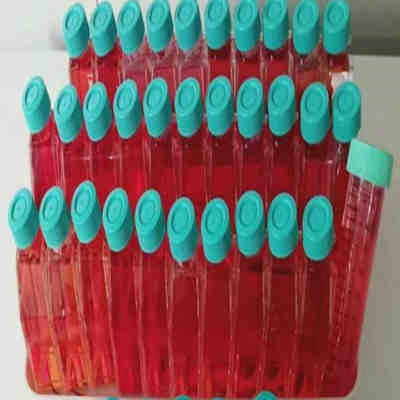
风险提示:丁香通仅作为第三方平台,为商家信息发布提供平台空间。用户咨询产品时请注意保护个人信息及财产安全,合理判断,谨慎选购商品,商家和用户对交易行为负责。对于医疗器械类产品,请先查证核实企业经营资质和医疗器械产品注册证情况。
 文献和实验
文献和实验模型主要分为三类:化学诱导模型、移植瘤模型和基因修饰自发肿瘤模型。 一、化学诱导模型 是指通过注射致癌物质如二甲基苯蒽等诱发小鼠乳腺癌。诱导模型构建简单,诱变剂选择范围大且稳定,诱发成癌率较高,类似于人体肿瘤细胞动力学特征,可用于乳腺癌预防及早期致癌因素的研究。但诱发模型肿瘤发生位点不可预知,同一动物可能有多个肿瘤出现,且肿瘤细胞形态学差异大,不适用于抗癌药物的研究。 二、移植瘤模型 将乳腺癌组织和细胞直接接种于实验动物而建立的模型,在基础研究中应用广泛。根据移植物来源可分为同种移植和异种移植,同种
-1 骨髓瘤 SRS-82 腹水瘤 SP2/0 骨髓瘤 SAC-IIB2 腹水瘤 P3-NS-1/1-Ag4.1 骨髓瘤 SAC-IIC3 腹水瘤 45.6.TG1.7 骨髓瘤 S180 腹水瘤 P3-X63-Ag8 骨髓瘤 B16 黑色素瘤 J774A.1 单核细胞-巨噬细胞 C127 乳腺肿瘤 RAW264.7 单核细胞-巨噬细胞 F9 胚胎瘤 NG108-15 小鼠神经细胞 瘤×大鼠神经胶质细胞杂交细胞
Nature 子刊:超声波调控肿瘤内细菌的基因表达,助力肿瘤免疫治疗
的潜在应用价值。 中科院深圳先进院合成所严飞研究员与南华大学附属第一医院陈智毅教授为共同通讯作者。深圳先进院客座学生陈宇浩和杜萌为共同第一作者。 由于肿瘤细胞的快速生长,许多实体肿瘤的内部经常会形成缺血、低氧、免疫抑制的肿瘤微环境,这大大限制了各种药物分子和免疫效应细胞在肿瘤内的渗透,成为各种恶性实体肿瘤难以根治的重要原因。幸运地是,实体肿瘤内部的这种低氧微环境也为肿瘤的细菌治疗提供了有利条件,某些厌氧菌和兼性厌氧菌能够靶向迁移到肿瘤核心低氧区,肿瘤内缺血和组织液压形成的免疫抑制微环境也为细菌
 技术资料
技术资料暂无技术资料 索取技术资料

