


- ¥3500 - 4500
- 欣润生物
- 江苏无锡
- IR3005-1
- 2026年04月28日

企业认证
相关产品推荐更多 >

万千商家帮你免费找货
0 人在求购买到急需产品
- 详细信息
- 文献和实验
- 技术资料
- 英文名:
A
- 库存:
10
- 供应商:
欣润生物
- 肿瘤类型:
NO
- 细胞类型:
永生化
- ATCC Number:
11222
- 品系:
SD大鼠
- 组织来源:
肝窦内皮
- 相关疾病:
无
- 物种来源:
大鼠
- 免疫类型:
不详
- 细胞形态:
上皮型
- 是否是肿瘤细胞:
否
- 器官来源:
/
- 运输方式:
常温
- 年限:
5年
- 生长状态:
贴壁生长
- 规格:
T25方瓶
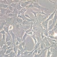
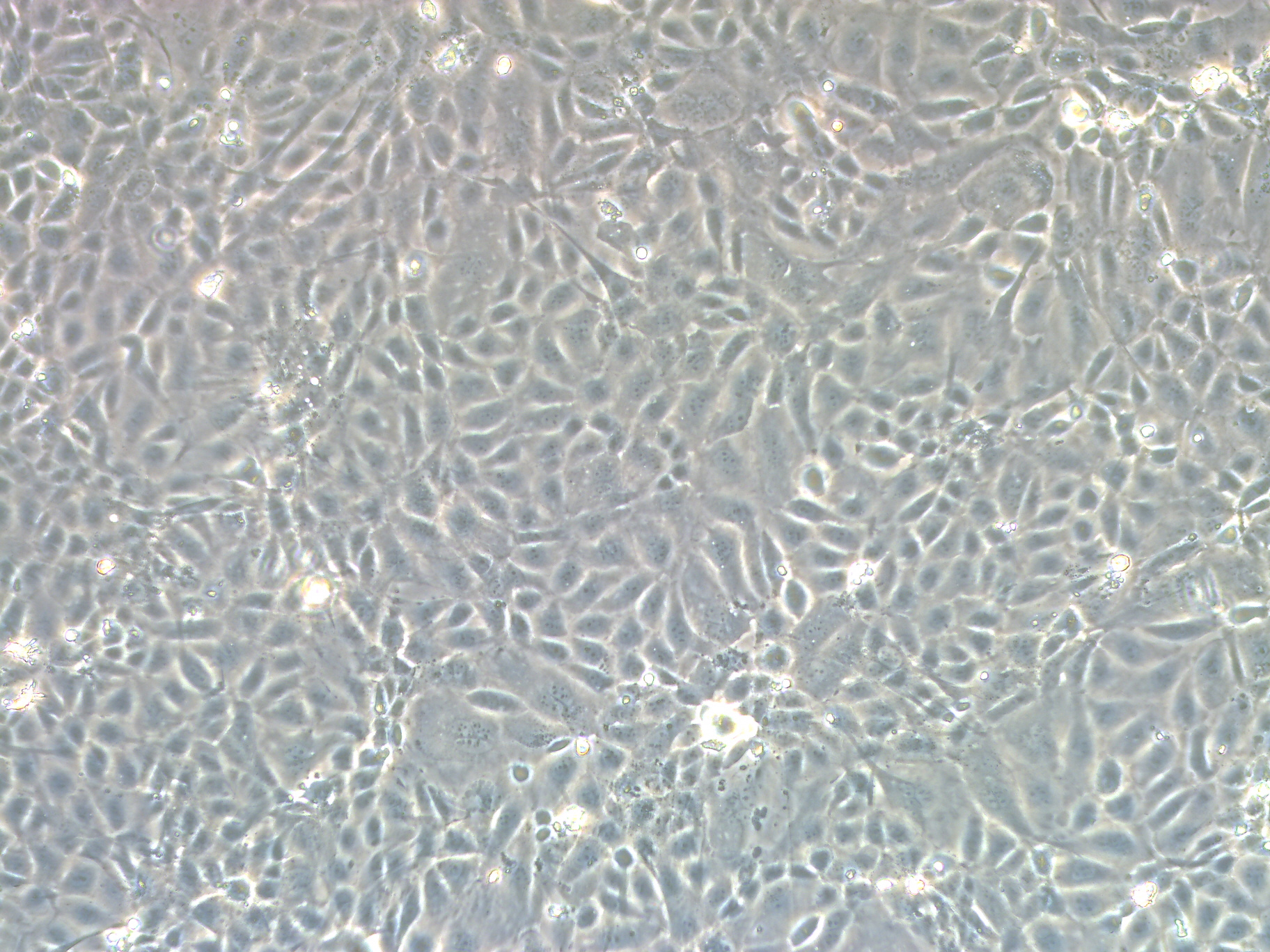
永生化大鼠肝窦内皮细胞简介:
虽然原代大鼠肝窦内皮细胞更能真实地反映肝窦内皮细胞在大鼠体内的生理状态,但是一方面原代分离培养肝窦内皮细胞周期长和对实验设备、试剂及技术人员经验要求较高,另一方面此原代细胞在体外的生长增殖能力非常缓慢有限,以至于此细胞不能传代,这些不利因素制约了原代大鼠肝窦内皮细胞在实验室的广泛应用。欣润生物的研究团队拥有多年原代细胞分离培养及细胞永生化服务研究经验,成功建立了永生化大鼠肝窦内皮细胞。
肝血窦是相邻肝板之间的腔隙,是一种特殊的毛细血管。肝血窦的窦壁由肝细胞的细胞膜构成,故肝血窦的通透性较大,有利于肝细胞与血流之间进行物质交换。在电镜下观察,肝血窦内皮细胞与肝细胞之间有一狭窄间隙,称窦周隙(Disse腔)。肝窦内皮细胞是肝脏非实质细胞的主要细胞群,由肝窦内皮细胞构成的肝窦壁是全身毛细血管壁中缺乏基膜的毛细血管窗孔,足肝窦内皮细胞最具特征性的结构。肝窦内皮细胞在调节肝窦血流与周围组织的物质交换中起有效的中枢性的作用,因此肝窦内皮细胞对于维持正常的肝功能起十分重要的作用。同时肝窦内皮细胞在肝脏的生理病理过程中发挥着诸多的重要功能。
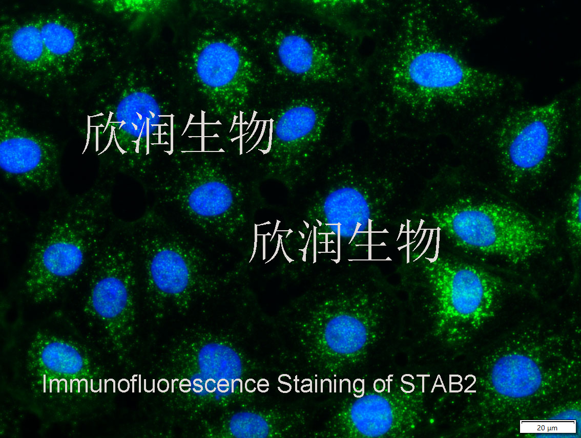
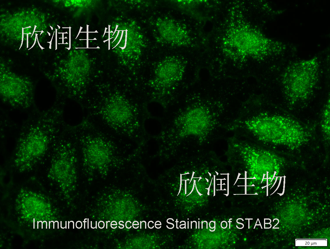
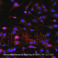
STAB2免疫荧光染色鉴定
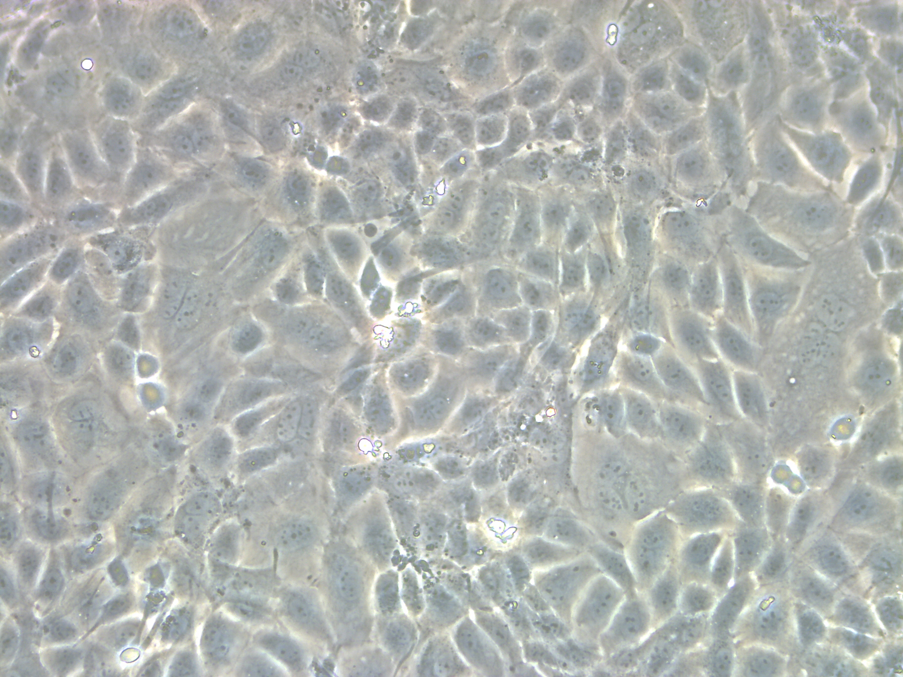
本公司生产的永生化大鼠肝窦内皮细胞采用体外灌注和密度梯度离心法和基因转染制备而来,细胞总量约为1×10⁶/T25方瓶,细胞纯度可达95%以上,且不含有HIV-1、 HBV、HCV、支原体、细菌、酵母和真菌等。
培养基信息:
我们推荐使用欣润生物研制的永生化大鼠肝窦内皮细胞完全培养基(产品编号:IR3005-5)作为体外培养永生化大鼠肝窦内皮细胞的培养基。
Study of the reappearance of sieve plate-like pores in immortalized sinusoidal endothelial cells – Effect of actin inhibitor in mixed perfusion cultures
Introduction We previously reported that when the high-functioning human hepatoma cell line, FLC-5, immortalized sinusoidal endothelial cell line, M1, and immortalized hepatic stellate cell line, A7, were cultured in the 3-dimensional filled type bioreactor, tissue reorganization resembling that seen in the live liver occurred, with the appearance of pores in the sinusoidal endothelial cells (SECs) [ 1 ]. The process and mechanism of formation of these pores remain unclarified. The presence of actin at the margin of these pores has been demonstrated by electron microscopic study [ 2 ]. Swinholide-A, which is actin inhibitor synthesized from Okinawa sponge, increase the number of pores on primary culture on SECs derived from the rat [ 3 ]. In present study, we examine whether or not the pores on SECs under three-dimensional perfusion co-culture treatment with Swinholide-A behave like those in primary culture cells. Methods 2 - 10 7 FLC-5 cells were inoculated into the reservoir, and they were per
Sphingosine 1-phosphate has anti-apoptotic effect on liver sinusoidal endothelial cells and proliferative effect on hepatocytes in a paracrine manner in human
Sphingosine 1-phosphate (S1P) is a bioactive sphingolipid metabolite released from erythrocytes and platelets, and is a potent stimulus for endothelial cell proliferation. However, the role of S1P on human liver sinusoidal endothelial cells (LSEC) remains unclear. The proliferation and inhibition of apoptosis in LSEC are involved in the promotion of liver regeneration and the suppression of liver injury after liver resection and transplantation. The aim of this study is to investigate the role of S1P on human LSEC and the interaction between S1P and LSEC in hepatocyte proliferation in vitro.Immortalized human LSEC were used. LSEC were cultured with S1P, and the cell proliferation, anti-apoptosis, signal transductions and production of cytokines and growth factors were subsequently examined. To investigate the interaction between S1P and LSEC in hepatocyte proliferation, primary human hepatocytes were cultured with the supernatants of LSEC with and without S1P. DNA synthesis and signal transductions in hepatocytes were examined.S1P induced LSEC proliferation through activation of Akt
风险提示:丁香通仅作为第三方平台,为商家信息发布提供平台空间。用户咨询产品时请注意保护个人信息及财产安全,合理判断,谨慎选购商品,商家和用户对交易行为负责。对于医疗器械类产品,请先查证核实企业经营资质和医疗器械产品注册证情况。
 文献和实验
文献和实验Chicken intestinal epithelial cells were obtained from NEWGAINBIO company. Cells were cultured on 37℃, with 5% CO2, in the Ham’s F-12 Nutrient (DMEM/12) that contained the following supplementations: fetal bovine serum (5%), in-sulin (5 µg/mL), transferrin (5 µg/mL), selenium (5 ng/mL), epidermal growth factor (5 ng/mL) and penicillin-streptomycin (100–100 U/mL) for cell culturing (full DMEM/12). Experiments were performed with chicken intestinal epithelial cells and working solutions were prepared with plain DMEM/12 without supplementation. For the investigations, cells were seeded onto 96-well, 24-well or 6-well polystyrene cell culture plates.
Primary hVICs (passage 2) were cultured to 50–60% confluence and infected with pGMLV-SV40T-puro lentivirus (NewgainBio, Wuxi, China) at a multiplicity of infection of 80 supplemented with 5 µg/mL polybrene (Sigma-Aldrich, Buchs, Switzerland).
Tissue was cultured until cells became visible around the tissue, and when the fusion reached 90% (FIGURE 1A) §ask ¦lled with the prepared culturing medium was sent to the company for further immortalisation. Cell immortalisation was done for cell stability and longer-term use. Immortalised cells were cultured with 10% FBS and 1% PS in the DMEM medium. After the cells multiplied and merged, they were routinely passed and grown ( NEWGAINBIO Inc. Wuxi, Jiangsu, China) (FIGURE 1B-C).
Mouse primary cultured renal vascular ECs and VSMCs were obtained from Newgainbio company, which were tested by Factor VIII and α-smooth muscle actin (α-SMA), the marker of ECs and VSMCs. RNeasy Mini Kit was used for RNA extraction, and the above protocols were repeated.
Porcine primary colon epithelial cells (Newgainbio company, Wuxi,China) were cultured in Dulbecco's Modified Eagle's Medium (Solarbio, Beijing, China) containing 10 % fetal bovine serum (BioInd, Kiryat shmona, Lsrael) at 37 ◦C and 5 % CO2 humidity.
的连接面有紧密连接、桥粒和缝隙连接等结构(图13-13)。 图13-13 肝细胞、肝血窦、窦周隙及胆小管的关系图解 图13-14 大鼠肝细胞电镜像 ×16100 N细胞核,RER粗面内质网,M线粒体,G高尔基复合体,Ly溶酶体,BC胆小管 (上海医科大学电镜室供图) 肝细胞核大而圆,居中央,常染色质丰富丰色浅,核膜清楚,核仁1至数个。部分肝细胞(约25%)有双核,有的肝细胞的核体积较大,为多倍体核。肝细胞核DNA含量分析,正常成体肝细胞
图13-19 大鼠肝电镜像示胆小管 ×30000 BC胆小管,HC肝细胞,↑紧密连接,()桥粒, ※高尔基复合体 (上海医科大学电镜室供图) 图13-20 大鼠肝冷冻割断扫描电镜像示胆小管 (河北医学院应国华教授供图) 图13-21 肝门管区 小叶间静脉:是门静脉的分支,管腔较大而不规则,壁薄,内皮外仅有少量散在的平滑肌。 小叶间动脉:是肝动脉的分支,管径较细,腔较小,管壁相对较厚,内皮外有几层环行平滑
图13-14 大鼠肝细胞电镜像 ×16100 N细胞核,RER粗面内质网,M线粒体,G高尔基复合体,Ly溶酶体,BC胆小管 (上海医科大学电镜室供图) 肝细胞核大而圆,居中央,常染色质丰富丰色浅,核膜清楚,核仁1至数个。部分肝细胞(约25%)有双核,有的肝细胞的核体积较大,为多倍体核。肝细胞核DNA含量分析,正常成体肝细胞以四倍体核占多数,约占肝细胞总数的70%左右,还有少量八倍体肝细胞。一般认为,双核肝细胞和多倍体肝细胞的功能比较活跃。肝细胞是一种高度分化并具有
 技术资料
技术资料
