


永生化大鼠肝Kupffer细胞、大鼠肝Kupffer细胞系、
大鼠Kupffer细胞系、永生化大鼠Kupffer细胞、大鼠肝库普弗细胞系、大鼠肝巨噬细胞系、永生化大鼠肝巨噬细胞- ¥3500 - 4500
- 欣润生物(NEWGAINBIO)
- 江苏无锡
- IR3003
- 2026年04月30日

企业认证
相关产品推荐更多 >


万千商家帮你免费找货
0 人在求购买到急需产品
- 详细信息
- 询价记录
- 文献和实验
- 技术资料
- 英文名:
Immortalized rat liver Kupffer cells
- 库存:
100万左右
- 供应商:
欣润生物
- 肿瘤类型:
否
- 细胞类型:
永生化
- ATCC Number:
无
- 品系:
SD
- 组织来源:
肝
- 相关疾病:
没有
- 物种来源:
大鼠
- 免疫类型:
不详
- 细胞形态:
不规则
- 是否是肿瘤细胞:
否
- 器官来源:
肝脏
- 运输方式:
常温
- 年限:
成年
- 生长状态:
贴壁生长
- 规格:
T25方瓶
细胞名称:永生化大鼠肝Kupffer细胞、大鼠肝巨噬细胞系、大鼠Kupffer细胞系、大鼠肝Kupffer细胞系、永生化大鼠肝库普弗细胞
细胞描述:永生化大鼠肝巨噬细胞系指位于肝脏肝窦内表面的巨噬细胞,它能够清除血液中的外原抗原及抗原-抗体复合物和细胞碎片等东西。永生化大Kupffer细胞系是全身单核-吞噬细胞系统的重要组成内容, 也是肝脏防御系统主要成员之一,在全身和肝脏疾病病历发生发展中起到重要的作用。
细胞产品货号:IR3003
细胞类型: 永生化细胞
细胞传代能力: 可以传代30代左右
细胞形态: 不规则形态
完全培养基:永生化大鼠肝Kupffer细胞完全培养基
支原体质控:检测呈阴性
细胞培养条件:37℃,5%CO2
发货方式:常温T25瓶子发货
货期:5-7天
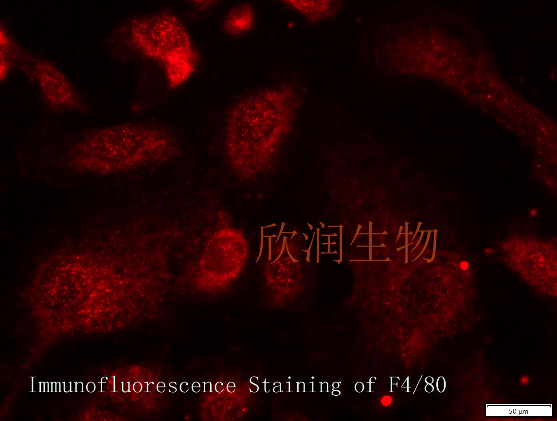
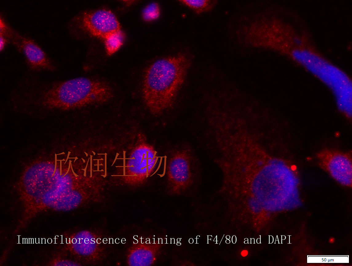
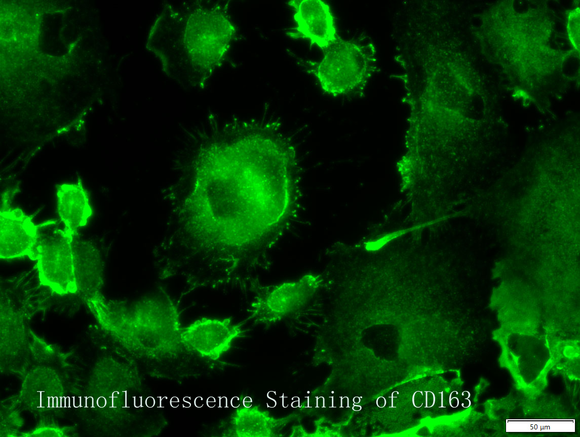

F4/80抗体和CD163抗体免疫荧光染色鉴定
引用文献:
GBP1 promotes acute rejection after liver transplantation by inducing Kupffer cells pyroptosis(2024)
期刊:BBA - Molecular Basis of Disease
DOI:10.1016/j.bbadis.2024.167644
影响因子:4.2


Establishment of immortalized rat Kupffer cell lines
Background. Kupffer cells have been implicated in the pathogenesis of various liver diseases. Primary cultures of Kupffer cells have a very limited life span, tend to de-differentiate and become senescent, and therefore are not suitable for cell signaling studies. Aim. To establish immortalized rat Kupffer cell lines that facilitate mechanistic studies of cell signaling and signal transduction. Methods. Rat Kupffer cells were sub-cultured with EGF to obtain rat Kupffer Cell line 1 (RKC1), and subsequently transfected with Simian Virus 40 Large T-antigen expression vector to obtain rat Kupffer Cell line 2 (RKC2). Results. RKC1 and RKC2 are similar to primary Kupffer cells as they express the molecular markers ED1, ED2, ED3, and F4/80, and upregulate TNF-α, IL-6, IL-1β, Fas /FasL, and NF-κB, as well as TLR4 in response to LPS or pancreatic elastase. Additionally, RKC1 and RKC2 maintain phagocytic properties of latex beads and exhibit increased telomerase and stabilized p53 activity. Conclusion. Immortalized RKC1 and RKC2 cells maintain properties of primary Kupffer cells and can be valuable tools in evaluating
Protocol for preparation of mouse liver Kupffer cells and liver sinusoidal endothelial cells
he following protocol is an outline of the method we use to prepare Kupffer cells (KC) and liver sinusoidal endothelial cells (LSEC) from mouse liver. The method is based on the one originally published to prepare rat liver KC and LSEC (1), and is slightly modified from the one outlined in (2). We also strongly recommend reading our 2012 review paper (3) to obtain a solid background understanding of the physiological functions of the LSEC.
Background Kupffer cells are well known macrophages of the liver, however, the developmental characteristics of Kupffer cells in mice are not well understood. To clarify this matter, the characteristics of Kupffer macrophages in normal developing mouse liver were studied using light microscopy and immunocytochemistry. Methods Sections of liver tissue from early postnatal mice were prepared using immunocytochemical techniques. The Kupffer cells were identified by their immunoreactivity to the F4/80 antibody, whereas endothelial cells were labelled with the CD-34 antibody. In addition, Kupffer cells and endothelial cells were labelled by systemically injected fluorescently labelled latex microspheres. Tissue slices were examined by fluorescence microscopy. Results Intravenous or intraperitonal injections of microspheres yielded similar patterns of liver cell labelling. The F4/80 positive Kupffer cells were labelled with both large (0.2 μm) and small (0.02 μm) diameter microspheres, while endothelial cells were labelled only with the smaller diameter microspheres. Microsphere
风险提示:丁香通仅作为第三方平台,为商家信息发布提供平台空间。用户咨询产品时请注意保护个人信息及财产安全,合理判断,谨慎选购商品,商家和用户对交易行为负责。对于医疗器械类产品,请先查证核实企业经营资质和医疗器械产品注册证情况。
- 作者
- 内容
- 询问日期
 文献和实验
文献和实验Chicken intestinal epithelial cells were obtained from NEWGAINBIO company. Cells were cultured on 37℃, with 5% CO2, in the Ham’s F-12 Nutrient (DMEM/12) that contained the following supplementations: fetal bovine serum (5%), in-sulin (5 µg/mL), transferrin (5 µg/mL), selenium (5 ng/mL), epidermal growth factor (5 ng/mL) and penicillin-streptomycin (100–100 U/mL) for cell culturing (full DMEM/12). Experiments were performed with chicken intestinal epithelial cells and working solutions were prepared with plain DMEM/12 without supplementation. For the investigations, cells were seeded onto 96-well, 24-well or 6-well polystyrene cell culture plates.
Primary hVICs (passage 2) were cultured to 50–60% confluence and infected with pGMLV-SV40T-puro lentivirus (NewgainBio, Wuxi, China) at a multiplicity of infection of 80 supplemented with 5 µg/mL polybrene (Sigma-Aldrich, Buchs, Switzerland).
Tissue was cultured until cells became visible around the tissue, and when the fusion reached 90% (FIGURE 1A) §ask ¦lled with the prepared culturing medium was sent to the company for further immortalisation. Cell immortalisation was done for cell stability and longer-term use. Immortalised cells were cultured with 10% FBS and 1% PS in the DMEM medium. After the cells multiplied and merged, they were routinely passed and grown ( NEWGAINBIO Inc. Wuxi, Jiangsu, China) (FIGURE 1B-C).
Mouse primary cultured renal vascular ECs and VSMCs were obtained from Newgainbio company, which were tested by Factor VIII and α-smooth muscle actin (α-SMA), the marker of ECs and VSMCs. RNeasy Mini Kit was used for RNA extraction, and the above protocols were repeated.
Porcine primary colon epithelial cells (Newgainbio company, Wuxi,China) were cultured in Dulbecco's Modified Eagle's Medium (Solarbio, Beijing, China) containing 10 % fetal bovine serum (BioInd, Kiryat shmona, Lsrael) at 37 ◦C and 5 % CO2 humidity.
canaliculi)等肝细胞结构,与肝脏天然组成类似,比2D培养更具功能性适用于特异质(idiosyncratic)和重复给药的长期毒性研究3.3D InSight™大鼠肝脏微组织(3D InSight™ Rat Liver Microtissues)即用型96孔板内大鼠肝微组织肝实质细胞与非实质细胞(non-parenchymal cells, NPCs)共培养,掺入了Kupffer细胞适用于特异质(idiosyncratic)和重复给药的长期毒性研究4.3D InSight™ HepG2肝脏微组织(3D
化镁);0.1mg/mL~1mg/mL的肝微粒体蛋白;合适浓度的待测物,一般推荐为1μM,于37°C水浴孵育,每个样品平行3次,以不包含NADPH发生系统的样品作为阴性对照。于预设的反应时间点,如0,5,10,15,30,60 min后加入等体积预冷的乙腈终止反应。 3.3 原型药物或代谢产物的检测 采用HPLC、HPLC-MC和HPLC-MC/MC测定温孵液中原型药物和其代谢产物。图5为采用LC-MS/MS测定药物在人、犬和大鼠肝微粒体中的代谢情况,从图上可知,药物在三种肝微粒体中均存在明显代谢,但不同
成纤维细胞仓鼠肾GMEM, 10% 胎牛血清 或MEM, 10% 胎牛血清 和NEAAHaK上皮细胞仓鼠肾BME, 10% 小牛血清CHO-K1上皮细胞仓鼠卵巢F-12, 10%胎牛血清AR42J.大鼠胰腺肿瘤Ham'sF-12K, 20%胎牛血清BRL3A.大鼠肝Coon'sF-12K, 5%胎牛血清Clone 9上皮细胞大鼠肝脏F-12K, 10%胎牛血清H4--Ⅱ-E-C3上皮样大鼠肝癌F-12K, 10%胎牛血清`GH1上皮细胞大鼠垂体肿瘤F-10, 15% 马血清和2.5% 胎牛血清GH
 技术资料
技术资料
