

- ¥308 - 528
- 翌圣生物(Yeasen)

- 40601ES
- 上海
- 2026年10月16日

企业认证
相关产品推荐更多 >
万千商家帮你免费找货
0 人在求购买到急需产品
相关阅读
源叶生物 十万种现货试剂速发,品质稳、品类全,实验不再等!Takara 秋促来袭,提取+反转录+定量套装仅 1200 元abinScience 体内级抗体 全年锁价 低至 ¥1200阿拉丁 22万+ 科研级产品现货速发,品质严控、科研无忧- 详细信息
- 文献和实验
- 技术资料
- 库存:
现货
- 供应商:
翌圣生物科技(上海)股份有限公司
- 英文名:
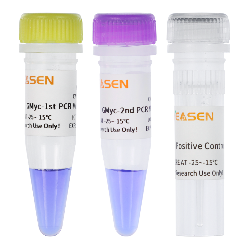
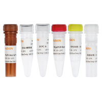
GMyc-PCR Mycoplasma Test Kit
- 规格:
10 assays(40601ES10)/20 assays(40601ES20)
| 规格: | 10 assays(40601ES10) | 产品价格: | ¥308 |
|---|---|---|---|
| 规格: | 20 assays(40601ES20) | 产品价格: | ¥528 |
产品描述:
细胞培养是生命科学研究中常见的实验。不像其它常用的实验方法,细胞培养是一个动态的连续过程,细胞通常会对操作失误或污染物有所反应,这些反应往往表现出不正常的细胞状态或培养基外观。如果受到支原体污染时,细胞形态较之无明显变化,极易被忽视,往往直到污染非常严重时才能发现。受污染的细胞膜上可能有几百个支原体,这些支原体竞争营养并释放有毒的代谢产物,严重影响实验结果。
研究表明,至少二十多种支原体能污染细胞,其中最常见的有:口腔支原体(M.orale)、精氨酸支原体(M.arginini)、猪鼻支原体(M.hyorhinis)、发酵支原体(M. fermentans)、人型支原体(M. hominis)、唾液支原体(M.salivarium)、肺支原体(M.pulmonis)和梨支原体(M.pirum)等。培养细胞的支原体污染率在4%-92%不等,其污染来源包括工作环境、操作者本身(一些支原体是人体的正常菌群)、培养基、血清、细胞交叉污染、实验器材和用来制备细胞的原始组织或器官的污染。
确认细胞培养过程中出现问题的潜在原因是一项困难且耗时的任务,在细胞培养中,任何突然的变化都应该被怀疑,同时良好的试验规范和定期检测支原体污染也十分必要。支原体检测的方法有很多种,如直接培养、DNA荧光染色、ELISA和PCR法等。
GMyc-PCR支原体检测试剂盒主要采用PCR方法对各种生物材料(如细胞培养物、实验动物分泌物、动物血清等)支原体感染的检测。它综合了几个优势:灵敏、特异、快速并且可以用直接用细胞培养上清液检测。本制品通过PCR方法检测培养细胞等生物材料中的支原体,所用引物为根据支原体16S-23S rRNA序列保守区域设计,只特异性扩增支原体DNA,检出灵敏度与特异性均较高。PCR扩增及电泳分析只需数个小时,操作方便简单。
产品特色:
1、灵敏度高:更低含量的支原体也能检测到,能够更早发现支原体污染
2、特异性强:和其他近亲缘菌样本同时检测,仅支原体被检出
3、适用范围广:可以用直接用细胞培养上清液检测
应用案例:
肺炎近缘生物菌 --无PCR扩增条带
| 英文名称 | 中文名称 |
| Acinetobacter_baumannii | 鲍曼不动杆菌 |
| Bacillus_subtilis | 枯草芽孢杆菌 |
| Clostridium_acetobutylicum | 有机溶剂丁醇梭菌 |
| Clostridium_perfringens | 产气荚膜梭菌 |
| Klebsiella_aerogenes | 产酸克雷伯氏菌 |
| Lactobacillus_acidophilus | 嗜酸乳杆菌 |
| Micrococcus_luteus | 藤黄微球菌 |
| Pseudomonas_aeruginosa | 铜绿假单胞菌 |
| Staphylococcus_epidermidis | 表皮葡萄球菌 |
| Streptococcus_mutans | 变异链球菌 |
| Streptococcus_pneumoniae | 肺炎链球菌 |
检测限更低10copies/μL
性能优于竞品
翌圣40601ES和竞品同时检测,40601特异性及灵敏度更高
储存条件:
干冰运输,-25~-15℃保存,有效期18个月。若较长时间不用,请避光保存。
风险提示:丁香通仅作为第三方平台,为商家信息发布提供平台空间。用户咨询产品时请注意保护个人信息及财产安全,合理判断,谨慎选购商品,商家和用户对交易行为负责。对于医疗器械类产品,请先查证核实企业经营资质和医疗器械产品注册证情况。
 文献和实验
1] Rao XS, Cong XX, Gao XK, et al. AMPK-mediated phosphorylation enhances the auto-inhibition of TBC1D17 to promote Rab5-dependent glucose uptake. Cell Death Differ. 2021;28(12):3214-3234. doi:10.1038/s41418-021-00809-9(IF:15.828)
[2] Guo F, Li L, Li J, et al. Single-cell multi-omics sequencing of mouse early embryos and embryonic stem cells. Cell Res. 2017;27(8):967-988. doi:10.1038/cr.2017.82(IF:15.606)
[3] Hao Y, He B, Wu L, et al. Nuclear translocation of p85β promotes tumorigenesis of PIK3CA helical domain mutant cancer. Nat Commun. 2022;13(1):1974. Published 2022 Apr 13. doi:10.1038/s41467-022-29585-x(IF:14.919)
[4] Shu X, Liu M, Lu Z, et al. Genome-wide mapping reveals that deoxyuridine is enriched in the human centromeric DNA. Nat Chem Biol. 2018;14(7):680-687. doi:10.1038/s41589-018-0065-9(IF:13.843)
[5] Li X, Xiong X, Wang K, et al. Transcriptome-wide mapping reveals reversible and dynamic N(1)-methyladenosine methylome. Nat Chem Biol. 2016;12(5):311-316. doi:10.1038/nchembio.2040(IF:12.709)
[6] Sun L, Yang X, Huang X, et al. 2-Hydroxylation of Fatty Acids Represses Colorectal Tumorigenesis and Metastasis via the YAP Transcriptional Axis. Cancer Res. 2021;81(2):289-302. doi:10.1158/0008-5472.CAN-20-1517(IF:12.701)
[7] Sun Z, Zhang Z, Wang QQ, Liu JL. Combined Inactivation of CTPS1 and ATR Is Synthetically Lethal to MYC-Overexpressing Cancer Cells. Cancer Res. 2022;82(6):1013-1024. doi:10.1158/0008-5472.CAN-21-1707(IF:12.701)
[8] Song J, Zhuang Y, Zhu C, et al. Differential roles of human PUS10 in miRNA processing and tRNA pseudouridylation. Nat Chem Biol. 2020;16(2):160-169. doi:10.1038/s41589-019-0420-5(IF:12.154)
[9] He B, Pan H, Zheng F, et al. Long noncoding RNA LINC00930 promotes PFKFB3-mediated tumor glycolysis and cell proliferation in nasopharyngeal carcinoma. J Exp Clin Cancer Res. 2022;41(1):77. Published 2022 Feb 24. doi:10.1186/s13046-022-02282-9(IF:11.161)
[10] Tang B, Liu BH, Liu ZY, Luo MY, Shi XH, Pang DW. Quantum Dots with a Compact Amphiphilic Zwitterionic Coating. ACS Appl Mater Interfaces. 2022;14(24):28097-28104. doi:10.1021/acsami.2c04438(IF:9.229)
[11] Huang C, Zhang Z, Chen L, et al. Acetylation within the N- and C-Terminal Domains of Src Regulates Distinct Roles of STAT3-Mediated Tumorigenesis. Cancer Res. 2018;78(11):2825-2838. doi:10.1158/0008-5472.CAN-17-2314(IF:9.130)
[12] Wu X, Yu M, Zhang Z, et al. DDB2 regulates DNA replication through PCNA-independent degradation of CDT2. Cell Biosci. 2021;11(1):34. Published 2021 Feb 8. doi:10.1186/s13578-021-00540-5(IF:7.133)
[13] Wang J, Zhang Y, Liu X, Liu H. Optimizing Adaptive Therapy Based on the Reachability to Tumor Resistant Subpopulation. Cancers (Basel). 2021;13(21):5262. Published 2021 Oct 20. doi:10.3390/cancers13215262(IF:6.639)
[14] Feng W, Liu R, Xie X, et al. SUMOylation of α-tubulin is a novel modification regulating microtubule dynamics. J Mol Cell Biol. 2021;13(2):91-103. doi:10.1093/jmcb/mjaa076(IF:6.216)
[15] Yu M, Hu X, Yan J, Wang Y, Lu F, Chang J. RIOK2 Inhibitor NSC139021 Exerts Anti-Tumor Effects on Glioblastoma via Inducing Skp2-Mediated Cell Cycle Arrest and Apoptosis. Biomedicines. 2021;9(9):1244. Published 2021 Sep 17. doi:10.3390/biomedicines9091244(IF:6.081)
[16] Ren S, Cai Y, Hu S, et al. Berberine exerts anti-tumor activity in diffuse large B-cell lymphoma by modulating c-myc/CD47 axis. Biochem Pharmacol. 2021;188:114576. doi:10.1016/j.bcp.2021.114576(IF:5.858)
[17] Wen F, Sun X, Sun C, et al. TAGLN Is Downregulated by TRAF6-Mediated Proteasomal Degradation in Prostate Cancer Cells. Mol Cancer Res. 2021;19(7):1113-1122. doi:10.1158/1541-7786.MCR-20-0513(IF:5.852)
[18] Tang B, Sun EZ, Zhang ZL, et al. Sphingomyelin-Sequestered Cholesterol Domain Recruits Formin-Binding Protein 17 for Constricting Clathrin-Coated Pits in Influenza Virus Entry. J Virol. 2022;96(5):e0181321. doi:10.1128/JVI.01813-21(IF:5.103)
[19] Hu J, Ren W, Qiu W, et al. Generation of induced pluripotent stem cell line (XDCMHi001-A) from an Ankylosing spondylitis patient with JAK2 mutation. Stem Cell Res. 2020;45:101788. doi:10.1016/j.scr.2020.101788(IF:4.495)
[20] Xiao S, Yao X, Ye J, Tian X, Yin Z, Zhou L. Epigenetic modification facilitates proline synthase PYCR1 aberrant expression in gastric cancer [published online ahead of print, 2022 May 30]. Biochim Biophys Acta Gene Regul Mech. 2022;1865(6):194829. doi:10.1016/j.bbagrm.2022.194829(IF:4.490)
[21] Wang J, Zhang Y, Liu X, Liu H. Is the Fixed Periodic Treatment Effective for the Tumor System without Complete Information?. Cancer Manag Res. 2021;13:8915-8928. Published 2021 Nov 30. doi:10.2147/CMAR.S339787(IF:3.989)
[22] Yang X, Ren S, Rehman ZU, et al. Molecular characterization, expression, and functional identification of TANK-binding kinase 1 (TBK1) of the cow (Bos taurus) and goat (Capra hircus). Dev Comp Immunol. 2022;133:104444. doi:10.1016/j.dci.2022.104444(IF:3.636)
[23] Zheng D, Chang X, Liu Y, et al. 2-Methoxy-5((3,4,5-trimethosyphenyl)seleninyl) phenol reverses EGF-induced cell migration and invasion through down-regulation of MDM2 in breast cancer cell lines. Cancer Biol Ther. 2019;20(4):513-523. doi:10.1080/15384047.2018.1537578(IF:3.373)
[24] Xu F, Zhang S, Liu Z, et al. TEX9 and eIF3b functionally synergize to promote the progression of esophageal squamous cell carcinoma. BMC Cancer. 2019;19(1):875. Published 2019 Sep 3. doi:10.1186/s12885-019-6071-9(IF:2.933)
[25] Pan H, Sun L, Wang W, et al. Serum long non-coding RNA LOC553103 as non-specific diagnostic and prognostic biomarker for common types of human cancer. Clin Chim Acta. 2020;508:69-76. doi:10.1016/j.cca.2020.05.017(IF:2.615)
[26] Li N, Lin SM, Li Y, Sun J, Zhang L, Chen M. An induced pluripotent stem cell line (GZHMCi004-A) derived from a fetus with heterozygous G380R mutation in FGFR3 gene causing achondroplasia. Stem Cell Res. 2021;53:102322. doi:10.1016/j.scr.2021.102322(IF:2.020)
[27] Luo Q, Wei C, Long Y, et al. Generation of an ELTD1 knockout human embryonic stem cell line by the iCRISPR/Cas9 system. Stem Cell Res. 2021;53:102350. doi:10.1016/j.scr.2021.102350(IF:2.020)
[28] Liu YQ, Ling TW, Wang HY, Yang YH, Song WJ, Wang TC. Generation of an integration-free induced pluripotent stem cell line (LZUSHI001-A) from an epileptic patient with DGKG mutation. Stem Cell Res. 2022;61:102768. doi:10.1016/j.scr.2022.102768(IF:2.020)
[29] Chen M, Lin SM, Li N, Li Y, Li Y, Zhang L. An induced pluripotent stem cell line (GZHMCi003-A) derived from a fetus with exon 3 heterozygous deletion in RUNX2 gene causing cleidocranial dysplasia. Stem Cell Res. 2021;51:102166. doi:10.1016/j.scr.2021.102166(IF:2.020)
[30] Xu Y, Wang X, Qiu T, et al. Generation of an induced pluripotent stem cell line (FDCHI007-A) derived from a patient with developmental and epileptic encephalopathy Type 31 carrying heterozygous c.545C > A mutation in DNM1 gene. Stem Cell Res. 2022;60:102709. doi:10.1016/j.scr.2022.102709(IF:2.020)
[31] Fan T, He J, Wang Y, Yu J, Sun W. Generation of an induced pluripotent stem cell line (FDCHi006-A) from a 7-year-old girl with central precocious puberty. Stem Cell Res. 2021;56:102542. doi:10.1016/j.scr.2021.102542(IF:2.020)
[32] Gong X, Zheng Z, Yang T, Zheng H, Xiao X, Jia N. Generation of an isogenic gene-corrected iPSC line (OGHFUi001-A-1) from a type 1 early infantile epileptic encephalopathy (EIEE1) patient with a hemizygous R330L mutation in the ARX gene. Stem Cell Res. 2022;60:102693. doi:10.1016/j.scr.2022.102693(IF:2.020)
[33] Jia N, Gong X, Chen J, et al. Generation of an induced pluripotent stem cell line (OGHFUi001-A) from a type 1 early infantile epileptic encephalopathy with ARX mutation. Stem Cell Res. 2021;53:102367. doi:10.1016/j.scr.2021.102367(IF:2.020)
[34] Zhu W, Zhou Y, Wang Q, et al. Generation of a human induced pluripotent stem cell (iPSC) line from skin fibroblasts of a patient carrying an E363Q mutation in PSEN1 gene. Stem Cell Res. 2022;61:102769. doi:10.1016/j.scr.2022.102769(IF:2.020)
[35] Luo F, Long K, Li X, et al. Deficient of LRRC8A attenuates hypoxia-induced necrosis in 3T3-L1 cells. Biosci Biotechnol Biochem. 2020;84(6):1139-1145. doi:10.1080/09168451.2020.1730689(IF:1.516)
你是如何确保该抗体在你的实验体系中发挥有效作用的,例如,是否使用同种型对照来确认抗体在你的细胞中的特异性?对这些方法的准确描述可以向读者展现出你的结果是值得信赖的。 一份检查清单 列一个检查清单,对撰写「材料与方法」部分十分有帮助。如下是一份基于前文信息的检查清单的例子: 细胞系:来源、目录号、认证,以及支原体检测的结果 动物:物种、品系、年龄、性别、饲养、来源,以及实验结束的方案 植物:来源、物种和品系、培养参数和储存条件 试剂和仪器设备:浓度、来源、目录号、制造商、型号,等等 抗体:目录号、来源
,支原体DNA会显示为特异性条带,以此指示支原体的存在。PCR法可以检测大多数支原体,但谨慎起见最好同时使用另一种检测方法来进行验证。 普健生物生产的支原体PCR检测试剂盒(Mycoplasma PCR Test Kit)是一款具有快速、灵敏、特异性高等特点的支原体检测试剂盒,可用于细胞、培养基、动物血清等的支原体检测。本产品通过聚合酶链式反应技术(PCR)特异扩增支原体16s-rRNA 基因保守区域片段进行支原体检测,能够检测包括M. arthritidis, M
对你的实验有用。 首先,细胞来源支原体污染的最常见原因之一是引入的培养物已经被感染。一旦进入实验室,这些小东西会蔓延到每个角落,让你崩溃。要避免这种情况,最简单的办法就是不从其他实验室要细胞,而从供应商或菌种保藏中心购买,这些细胞经过严格检测,确保不含有支原体。如果购买有难度,或基于其他原因,不得不使用其他实验室的细胞,那么也要保持警惕,在单独的培养箱中隔离,同时进行支原体的检测。市场上有多家公司提供支原体检测试剂盒,你可自行选择。 其次,定期检测即使实验室没有新来的细胞,支原体也可能通过操作