大家都在搜

手机验证

询价列表

暂时没有已询价产品
bioyxs
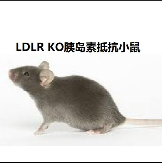
背景:
应用LDL受体(低密度脂蛋白受体,LDLR)敲除的小鼠(LDLR-/-小鼠)研究胰岛素抵抗有很多优点和特殊性,例如:
(1) 胰岛素抵抗容易被诱导,有利于药物或保健食品对胰岛素抵抗作用的筛选和效果观察;
(2) 在高脂模型饲料喂养后很快发生血浆胰岛素水平升高、血糖升高(糖尿病),因此,LDLR-/-小鼠是胰岛素抵抗向II型糖尿病发展的理想模型;
(3) LDLR-/-小鼠的血脂和胆固醇分别非常接近人类,在该小鼠中研究胰岛素抵抗与血脂改变的关系,比在大鼠和普通小鼠中研究有独特的优势。
(4) 高脂模型饲料喂养后LDLR-/-小鼠容易发生肥胖、非酒精性脂肪肝和动脉粥样硬化,在该小鼠中研究胰岛素抵抗有利于分析胰岛素抵抗与这些疾病的关系。
LDLR-KO小鼠在喂养高脂模型饲料后的几周内发生胰岛素抵抗、高胰岛素血症,并且很快发生高血糖。根据研究报道,胰岛素抵抗发生后持久维持。但相继发生脂肪肝和动脉粥样硬化,因此,应当根据你的研究具体情况确定最合适的造模时间。
(5) 研究胰岛素抵抗与LDLR的功能关系
Atherosclerosis measurement in male WT → Ldlr KO and Lpcat3 KO → Ldlr KO mice. (A) Aortic arches with atherosclerotic plaques (white areas). (B) Aortic root assay for lesion areas after H&E staining. Six alternate sections (10 μm thick) sliced from paraffin-fixed aortic root tissues of each transplanted mouse were used for the analysis. (C) En face aortic plaque analysis after Oil Red O staining. (D) Quantitative display of root assay. (E) Quantitative display of en face assay. Red arrows indicate lesion area. Values are mean ± SD. N = 8–9.
风险提示:丁香通仅作为第三方平台,为商家信息发布提供平台空间。用户咨询产品时请注意保护个人信息及财产安全,合理判断,谨慎选购商品,商家和用户对交易行为负责。对于医疗器械类产品,请先查证核实企业经营资质和医疗器械产品注册证情况。
 询价记录
询价记录