

- ¥1480
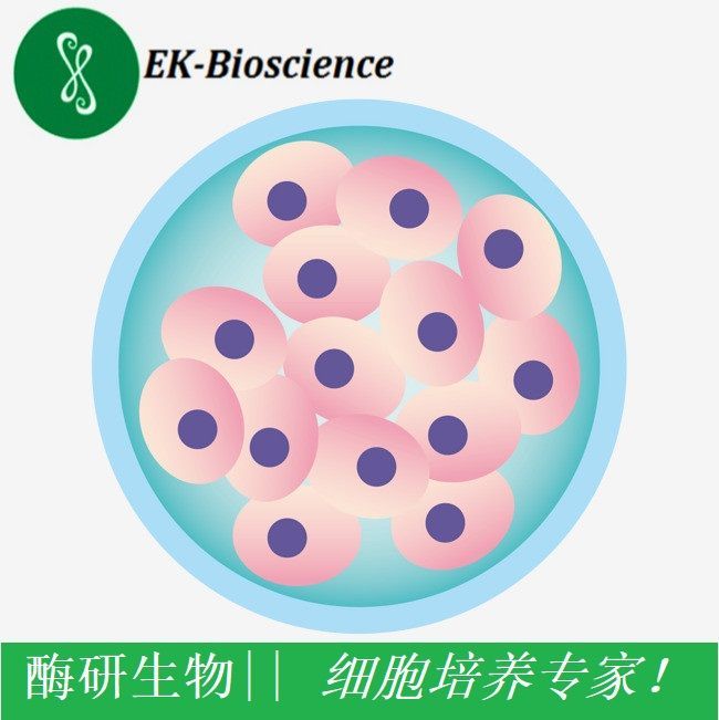
- EK-Bioscience

- 国内
- MY-K5284
- 2026年01月04日

企业认证
相关产品推荐更多 >

万千商家帮你免费找货
0 人在求购买到急需产品
- 详细信息
- 文献和实验
- 技术资料
- 英文名:
sk-hep1
- 库存:
1x10^6/瓶/支
- 供应商:
上海酶研
- 肿瘤类型:
详询
- 细胞类型:
人肝癌细胞
- ATCC Number:
详询
- 品系:
sk-hep1
- 组织来源:
人肝癌细胞
- 相关疾病:
sk-hep1
- 物种来源:
哺乳动物
- 免疫类型:
详询
- 细胞形态:
贴壁/悬浮
- 是否是肿瘤细胞:
详询
- 器官来源:
人肝癌细胞
- 运输方式:
顺丰快递
- 年限:
5年
- 生长状态:
生长良好
sk-hep1 /sk-hep1 细胞系/sk-hep1 细胞株/sk-hep1 人肝癌细胞
Cell line name SK-HEP-1
Synonyms SK-Hep-1; SK HEP-1; SK HEP 01; SK-Hep1; Sk-Hep1; SK Hep1; SKHEP-1; SKHEP1; SKHep1; SK_HEP1
Accession CVCL_0525
Resource Identification Initiative To cite this cell line use: SK-HEP-1 (RRID:CVCL_0525)
Comments Problematic cell line: Misclassified. Originally described as originating from an adenocarcinoma of liver and thus classified as hepatocellular carcinoma. Later studies show that it most probably has arisen from endothelial cells (PubMed=1371504; PubMed=31938418).
Part of: Cancer Dependency Map project (DepMap) (includes Cancer Cell Line Encyclopedia - CCLE).
Part of: COSMIC cell lines project.
Part of: MD Anderson Cell Lines Project.
From: Memorial Sloan Kettering Cancer Center; New York; USA.
Registration: Memorial Sloan Kettering Cancer Center Office of Technology Development; SK1980-535.
Population: Caucasian.
Doubling time: 46.7 +- 10.3 hours, 94.2 hours (Note=In CDM4-CHO medium), 289.8 hours (Note=In 293 SFM II medium) (PubMed=25822314); ~30 hours (DSMZ=ACC-141).
Karyotypic information: Has lost chromosome Y.
Microsatellite instability: Stable (MSS) (Sanger).
Omics: Deep exome analysis.
Omics: Deep quantitative proteome analysis.
Omics: DNA methylation analysis.
Omics: Protein expression by reverse-phase protein arrays.
Omics: Secretome proteome analysis.
Omics: SNP array analysis.
Omics: Transcriptome analysis by microarray.
Omics: Transcriptome analysis by RNAseq.
Derived from site: Metastatic; Ascites; UBERON=UBERON_0007795.
Cell type: Endothelial cell; CL=CL_0000115.
PubMed=3518877; DOI=10.3109/07357908609038260
Fogh J.
Human tumor lines for cancer research.
Cancer Invest. 4:157-184(1986)
PubMed=2439335; DOI=10.1111/j.1432-1033.1987.tb11497.x
Vincent C., Marceau M., Blangarin P., Bouic P., Madjar J.-J., Revillard J.-P.
Purification of alpha 1-microglobulin produced by human hepatoma cell lines. Biochemical characterization and comparison with alpha 1-microglobulin synthesized by human hepatocytes.
Eur. J. Biochem. 165:699-704(1987)
PubMed=1371504; DOI=10.1007/BF02631017
Heffelfinger S.C., Hawkins H.H., Barrish J., Taylor L., Darlington G.J.
SK HEP-1: a human cell line of endothelial origin.
In Vitro Cell. Dev. Biol. Anim. 28:136-142(1992)
PubMed=8224613; DOI=10.1096/fasebj.7.14.8224613
Puisieux A., Galvin K., Troalen F., Bressac B., Marcais C., Galun E., Ponchel F., Yakicier C., Ji J.-W., Ozturk M.
Retinoblastoma and p53 tumor suppressor genes in human hepatoma cell lines.
FASEB J. 7:1407-1413(1993)
PubMed=8389256; DOI=10.1093/carcin/14.5.987
Hsu I.C., Tokiwa T., Bennett W., Metcalf R.A., Welsh J.A., Sun T., Harris C.C.
p53 gene mutation and integrated hepatitis B viral DNA sequences in human liver cancer cell lines.
Carcinogenesis 14:987-992(1993)
PubMed=9023415; DOI=10.1006/cimm.1996.1062
Seki N., Hoshino T., Kikuchi M., Hayashi A., Itoh K.
HLA-A locus-restricted and tumor-specific CTLs in tumor-infiltrating lymphocytes of patients with non-small cell lung cancer.
Cell. Immunol. 175:101-110(1997)
PubMed=9178645; DOI=10.1006/cimm.1997.1108
Nakao M., Sata M., Saitsu H., Yutani S., Kawamoto M., Kojiro M., Itoh K.
CD4+ hepatic cancer-specific cytotoxic T lymphocytes in patients with hepatocellular carcinoma.
Cell. Immunol. 177:176-181(1997)
PubMed=11416159; DOI=10.1073/pnas.121616198; PMCID=PMC35459
Masters J.R.W., Thomson J.A., Daly-Burns B., Reid Y.A., Dirks W.G., Packer P., Toji L.H., Ohno T., Tanabe H., Arlett C.F., Kelland L.R., Harrison M., Virmani A.K., Ward T.H., Ayres K.L., Debenham P.G.
Short tandem repeat profiling provides an international reference standard for human cell lines.
Proc. Natl. Acad. Sci. U.S.A. 98:8012-8017(2001)
PubMed=12029633; DOI=10.1053/jhep.2002.33683
Yasui K., Arii S., Zhao C., Imoto I., Ueda M., Nagai H., Emi M., Inazawa J.
TFDP1, CUL4A, and CDC16 identified as targets for amplification at 13q34 in hepatocellular carcinomas.
Hepatology 35:1476-1484(2002)
风险提示:丁香通仅作为第三方平台,为商家信息发布提供平台空间。用户咨询产品时请注意保护个人信息及财产安全,合理判断,谨慎选购商品,商家和用户对交易行为负责。对于医疗器械类产品,请先查证核实企业经营资质和医疗器械产品注册证情况。
 文献和实验
文献和实验*发表【中文论文】请标注:由上海酶研生物科技有限公司提供;
*发表【英文论文】请标注:From Shanghai EK-Bioscience Biotechnology Co., Ltd.
that may be useful in the analysis of real-time, quantitative PCR data. 全文地址: http://www.sciencedirect.com/science?_ob=MImg&_imagekey=B6WN5-457MF4S-3-1&_cdi=6953&_user=554529&_orig=search&_coverDate=12%2F31%2F2001&_qd=1&_sk=999749995&view=c&wchp=dGLbVlz-zSkzS&md
Sucrose Density Gradient Fractionation
about 1Add 1 ml of COLD 1M NaN3 to 100 ml culture (we want 10 mM total conc.)Spin down 3 x10⁹ cells (about 100 O.D.equivalents (100 ml of OD = 1): 2750 X g.,10 min.,30℃ in lab clinical centrifugeWash 1x with 10 ml SK buffer in 15 ml falcon tubes (see recipe
Sucrose Density Gradient Fractionation of Yeast Membranes
to conserve zymolyase--it's expensive) Shake gently, 45 min, 60 rpm, 30oC FROM HERE ON OUT KEEP EVERYTHING AT 4oC Centrifuge 500 x g, 10 min. 4oC. Wash 1 x with 2 ml cold SK buffer Wash again with 2 ml lysis buffer C
 技术资料
技术资料
