

- 询价
- Biotium

- 美国
- 30050
- 2025年07月16日


相关产品推荐更多 >

万千商家帮你免费找货
0 人在求购买到急需产品
- 详细信息
- 文献和实验
- 技术资料
- 保存条件:
-20°C
- 保质期:
6个月
- 英文名:
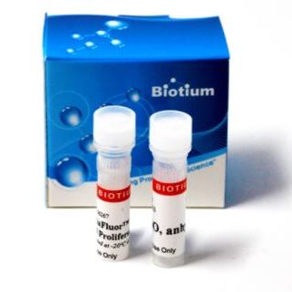
ViaFluor CFSE Cell Proliferation Kit 30050
- 库存:
99
- 供应商:
Biotium
- CAS号:
1
ViaFluor® SE Cell Proliferation Kits use amine-reactive dyes to covalently label cells throughout the cell cytoplasm and intracellular compartments for fixable fluorescent staining. Cell proliferation dyes are commonly used to monitor cell division by flow cytometry. The dyes also can be used to stably label cells to image cell morphology, or to track cell populations in mixed co-culture experiments.
Features
- Non-toxic dyes covalently label cell cytoplasm for fixable staining
- Track cell proliferation in vivo or in vitro by dye dilution using flow cytometry
- Long term imaging of cell morphology or co-cultures by microscopy
- Excellent performance & lower cost compared to leading competitors
- ViaFluor® 405 & ViaFluor® 488 SE are much less toxic than CFSE
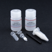
Kit Components
- Lyophilized dye in single use vials
- Anhydrous DMSO for dissolving dye
Spectral Properties (Ex/Em after hydrolysis)
- ViaFluor® CFSE: 495/515 nm
- ViaFluor® 488 SE: 493/532 nm
- ViaFluor® 405 SE: 408/452 nm
ViaFluor® SE Dyes for Cell Proliferation Tracking
ViaFluor® SE dyes are membrane-permeant compounds that are initially non-fluorescent esters, but are converted to fluorescent dyes by intracellular esterases and will covalently react with amine groups on intracellular proteins at the same time, forming fluorescent conjugates that are retained in the cell. Immediately after staining, a single bright fluorescent population will be detected by flow cytometry. With each cell division, daughter cells inherit roughly half of the fluorescent label, allowing the number of cell divisions that occur after labeling to be detected by the appearance of successively dimmer fluorescent peaks on a flow cytometry histogram compared to cells analyzed immediately after staining. Thus, cell proliferation dyes can be used to track multiple cell divisions of cells grown in culture or injected in vivo after labeling with the ViaFluor® SE dye.
The number of assays that can be performed per kit depends on the dye concentration used (see the product protocol for more information). When used at 1 uM to label 106 cells in one mL, each dye vial can be used for 90-100 labelings.
Other Applications for ViaFluor® SE Dyes
ViaFluor® SE dyes also can be used for imaging cell morphology or identifying cells in co-culture by microscopy. Because the staining is non-toxic and well-retained, it can be used for imaging live cells over time. See our Tech Tip: Using ViaFluor® SE Stains for Cell Tracing and Co-Culture.
All three ViaFluor® SE dyes can stain gram-positive bacteria, but not gram-negative bacteria. ViaFluor® CFSE stains the cytoplasm in yeast, but ViaFluor® 405 & ViaFluor® 488 stain the yeast cell periphery. See our Cellular Stains Table for more information on how our dyes stain various organisms.
Cell Division |
Catalog No. |
Ex/Em (nm) |
Flow detection |
Features |
|---|---|---|---|---|
| ViaFluor® 405 Cell Proliferation Kit | 30068 | 408/452 | Pacific Blue® | • Track cell division by dye dilution using flow cytometry • ViaFluor® 488 is a unique, improved green dye to replace CFSE • ViaFluor® 405 replaces CellTrace™ Violet |
| ViaFluor® 488 Cell Proliferation Kit | 30086 | 493/532 | FITC | |
| ViaFluor® CFSE Cell Proliferation Kit | 30050 | 495/519 | FITC |
风险提示:丁香通仅作为第三方平台,为商家信息发布提供平台空间。用户咨询产品时请注意保护个人信息及财产安全,合理判断,谨慎选购商品,商家和用户对交易行为负责。对于医疗器械类产品,请先查证核实企业经营资质和医疗器械产品注册证情况。
 文献和实验
文献和实验细胞仪检测分析,通过检测到细胞荧光强度不断的降低,进一步分析得出细胞分裂增殖的情况。二、CFSE配制用DMSO溶解成5 mmol/L的储存液,于- 20 ℃避光保存。使用时,用无血清DMEM培养液稀释成5μmol/L的工作液备用。CFSE试剂盒内有一小瓶CFSE(A试剂)标明500ug ,另有一小瓶DMSO(B试剂),500ugCFSE溶解于180ulDMSO即是5mMstock solution。三、CFSE标记细胞制作细胞悬液,加入等体积CFSE工作液,于37℃孵育10 min,用40%体积的冷小牛血清立即终止
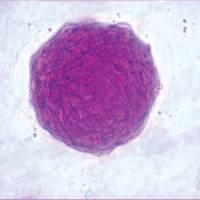
检测细胞增殖的方法很多,MTT、CCK8、BrdU 等等,你知道细胞可以 CFSE 染色用流式来检测细胞增殖么?好啦,不知道的同学搬好小板凳排排坐啦!CFSE 是一种荧光染料,脂溶性高,易进入细胞,可以与胞内氨基酸发生不可逆的结合,在细胞的分裂过程中能够进入子代细胞,同时荧光强度也变为母细胞的一半,可用流式或荧光显微镜对其进行分析。下面我们就开始动手,用流式来检测脾淋巴细胞的增殖。☟☟☟— 先准备一些试剂 —ConA 储存液:用双蒸水配置终浓度为 1 mg/ml(200×)CFSE 储存
一、胸腺嘧啶核苷(3H-TdR)渗入法胸腺嘧啶核苷(TdR)是DNA特有的碱基,也是DNA合成的必需物质。用同位素3H标记TdR即3H-TdR作为DNA合成的前体能掺入DAN合成代谢过程,通过测定细胞的放射性强度,可以反映细胞DAN的代谢及细胞增殖情况。但是具有放射性。二、MTT检测法MTT检测法主要反映细胞的能量代谢,是检测细胞增殖活力的一种简便准确的方法,其原理是在活细胞生长和增殖过程中,线粒体内的脱氢酶可将黄色的MTT分解成兰紫色的甲(Formazan),生成的甲量的多少与细胞的数量


