Imaging Surface and Submembranous Structures in Living Cells With the Atomic Force Microscope: Notes and Tricks
互联网
互联网
相关产品推荐

p46/p46蛋白Recombinant Mesomycoplasma hyopneumoniae 46 kDa surface antigen (p46)重组蛋白p46蛋白
¥2616

PerCP-Cy5.5 Anti-Mouse CD49b/pan-NK cells Antibody(DX5)
¥1890

噻唑蓝四唑蓝,298-93-1,Membrane-permeable yellow dye that is reduced by mitochondrial reductases in living cells to form the dark blue product, MTT-formazan.,阿拉丁
¥895.90

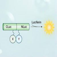
荧火素酶互补实验(Luciferase Complementation Assay, LCA)| 荧光素酶互补成像技术(Luciferase Complementation Imaging, LCI)
¥5999

pal/pal蛋白Recombinant Legionella pneumophila Peptidoglycan-associated lipoprotein (pal)重组蛋白19KDA surface antigen PPL蛋白
¥2328
相关问答

