Imaging Cellular and Molecular Dynamics in Live Embryos Using Fluorescent Proteins
互联网
互联网
相关产品推荐
Recombinant-Hordeum-vulgare-High-molecular-mass-early-light-inducible-protein-HV58-chloroplasticHigh molecular mass early light-inducible protein HV58, chloroplastic; ELIP
¥10556
yscM/yscM蛋白/yscM; Yop proteins translocation protein M蛋白/Recombinant Yersinia enterocolitica Yop proteins translocation protein M (yscM)重组蛋白
¥69
CSE1L/CSE1L蛋白Recombinant Human Exportin-2 (CSE1L)重组蛋白Cellular apoptosis susceptibility protein Chromosome segregation 1-like protein Importin-alpha re-exporter蛋白
¥5268
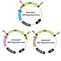
NLRP5 Homo sapiens maternal-antigen-that-embryos-require protein (MATER) mRNA.
询价
RMDN3/RMDN3蛋白Recombinant Human Regulator of microtubule dynamics protein 3 (RMDN3)重组蛋白Cerebral protein 10Protein FAM82A2;Protein FAM82;CProtein tyrosine phosphatase-interacting protein 51TCPTP-interacting protein 51蛋白
¥1344
相关问答

