UV Shadowing
互联网
UV shadowing is a technique for visualizing nucleic acids separated on acrylamide/urea gels. The technique utilizes shortwave UV light (254 nm) and a fluor-coated TLC plate. We recommend UV shadowing as the method of choice for gel purification of nonisotopic probes synthesized with Ambion's MAXIscript™ and BrightStar™ Psoralen-Biotin Kits. The alternative to UV shadowing is staining with ethidium bromide or acridine orange and requires subsequent extraction of the dye. The detection limit of UV shadowing is approximately 0.3 µg of nucleic acid.
Procedure
1.After electrophoresis, remove one of the glass plates and cover the gel with plastic wrap.
2.Place the gel, plastic wrap-side-down, on a flat surface and slowly remove the other glass plate.
3.(optional) Cover the gel with a second sheet of plastic wrap.
4.In a darkened room, place the plastic-wrapped gel on top of the fluor-coated TLC plate.
5.Visualize nucleic acid bands by shining a hand-held UV light source (254 nm; shortwave) on the surface of the gel.
6.Nucleic acid will appear as dark purple bands while the TLC plate will appear green.
Note: The xylene cyanol and bromophenol blue dyes present in most gel loading buffers may also appear as dark bands during UV shadowing and are sometimes mistaken for nucleic acid. We recommend that you run an extra lane of loading buffer alone in order to easily distinguish between these dyes and a shadow caused by nucleic acids.
7.Mark the band to be removed.
8.Cut out and remove the band corresponding to the desired nucleic acid.

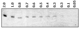
Figure 1. Microgram Dilutions (as Labeled) of an RNA Transcript Separated on a Denaturing 0.75 mm Thick 5% Acrylamide/8 M Urea Gel. The well dimensions were 6 mm wide. After electrophoresis, the gel was removed from the glass plates and UV shadowed from above with hand-held, shortwave UV source. The image was captured on an Alpha Innotech 500 imaging system.