免疫组化技术-Immunohistochemical Protocols
互联网
National Institute of Neurological Disorders and Stroke and Béla Hunyady
National Institute of Mental Health,National Institutes of Health
Bethesda, Maryland 20892
http://intramural.nimh.nih.gov/lcmr/snge/Protocols/IHH/immuno.html
Both immunohistochemistry (IHC) and
in situ hybridization histochemistry (ISHH)
are used to localize proteins (peptides) and mRNA to cells. No other methods give cellular resolution within tissues. Colocalization of gene expression can be gained by double IHC (colocalizing two different antigens in the same cells), by double
ISHH
(colocalizing two mRNAs in the same cells), and by the combination of the two methods (colocalizing an antigen and an mRNA in the same cells). Below are protocols that we are routinely using in our laboratory.
The presence of reagents and their sources in these protocols does not mean that there are no other alternatives, of course. The list does not represent an endorsement in any way by the U.S. Government.
The principle of immunohistochemistry is to raise an antibody to a specific antigen and use it as a marker to search for the antigen in tissue. The antibody can be directly conjugated to a fluorochrome or biotin that can then be detected in several different ways. Much more common, however, is the use of a non-conjugated primary antibody (mostly these are IgG's), and then an appropriate pre-conjugated anti-IgG molecule to detect the primary antibody. In addition to these conventional immunostainings, a newer method has been introduced with increased sensitivity. This is called the tyramide signal amplification (TSA) method. The permutations of the technique are so broad that it is not feasible even to try to describe all the possible immunostaining pathways (see table 1 ). We describe below some basic protocols that we have used and found successful.
Slides for mounting tissue sections:
-
Subbed slides
-
Place slides in racks and soak in soap solution for 1 hr.
-
Rinse in deionized water. Change the water several times to be sure that all of the soap is removed.
-
Dissolve 1.88 g of gelatin (300 bloom swine) in 750 ml hot H
2
O (do not allow to boil). Cool and dissolve 0.188 g CrK(SO
4
)
2
~undefined12H
2
O in the solution.
-
Dip the slides into the subbing solution, drain the slides onto a paper towel and allow to air dry for 1 hr.
-
Dip the slides into the subbing solution again. Drain and cover loosely with plastic wrap or bench paper.
- When thoroughly dry, store the slides in slide boxes.
-
Silanized slides:
-
Clean slides with a lint free cloth manually (lots of work, but needed) and put them in racks.
-
Dip slides in 2% aminoalkalynsilane (Sigma A-3648) in dry acetone for 10 sec.
-
Rinse in deionized water 3 times.
- Air dry overnight and store in boxes protected from dust.
-
Positively charged slides:
-
We buy these slides: Superfrost Plus microscope slides (4951+, Erie Scientific, Portsmouth, NH 03801) or you can make them as follows:
-
Clean slides with a lint free cloth manually (lots of work, but needed) and put them in racks
Dip slides in 50?g/ml poly-L-lysine
- Air dry overnight and store in boxes protected from dust.
Tissue preparation:
Two kinds of tissue samples are used. Fresh frozen tissues are mounted on tissue holders and 12µm sections are cut in a cryostat. Or, alternatively, animals are perfused through the ascending aorta under pentobarbital anesthesia with paraformaldehyde, as follows: The chest of the anesthetized animal is opened, and the ascending aorta cannulated through the left ventricle of the heart. After opening the left atrium, the vasculature is washed with 100 ml of 0.9 % NaCl (37°C) for five min, using a perfusion pump (Ismatec MV-CA, Zurich, Switzerland). The animals are then perfused with 500 ml 4% paraformaldehyde fixative over 20 min. The organs are removed and postfixed in the same fixative for 30 min. Subsequently, the tissues are cryoprotected in 5% (2 h), 10 % (4 h) and 20% (overnight, 4°C) sucrose in 1x PBS (pH 7.4) solutions, and frozen in 2-methylbutane (Aldrich, Milwaukee, WIS #M3,263-1) or on powdered dry ice. There are advantages and disadvantages to both tissue preparations: perfusion needs a special set-up, special expertise and is more time consuming. However, most antigens are well-preserved this way and also the tissue structure is excellent. Immersing the fresh frozen sections into fixative (mostly formaldehyde) is fast, easy but preservation of antigenicity and tissue structure is less good. However, immersion fixation in the non-crosslinking Histochoice (Amresco) may provide a good alternative. For some tissues there is no perfusion alternative (human biopsies or postmortem tissues), so that immersion fixation is the only way to go.
In general, sections from fresh frozen tissue are immersed into 4% formaldehyde (in 1xPBS) for 10 min at room temperature, and afterwards are treated identically to sections of perfused tissue. A blocking step is necessary to prevent non-specific binding of the primary antibody. For example if the secondary antibody is made in goat, than anything that would bind goat IgG should be blocked prior to staining. This goal is achieved by a preincubation (=blocking) with 5% normal goat serum in PBS. Blocking can also be achieved by using bovine serum albumine (BSA) instead of NGS. The solution should also contain 0.6% Triton X-100 to improve penetration into the tissue.
One needs to decide in advance if the end result (final marker or tag) should be fluorescent or suitable for regular brightfield microscopy (e.g., DAB reaction).
See below for composition of Reagents
Immunohistochemical methods for staining sections on slides:
(Summaries of each of the immunohistochemical paths below are presented in
table 1
)
-
The frozen sections are fixed in 4% paraformaldehyde fixative for ten min, followed by washes in PBS.
-
To decrease nonspecific staining, the fixed sections are incubated in BSA diluent for 30 min, and washed in PBS.
-
The schematic protocols of the applied immunohistochemical procedures are summarized in Table 1. We refer to the different immunostaining paths by the numbers in italics according to this table.
After the immunostainings, described in the next paragraphs, the sections are washed in distilled water, air dried, and coverslipped using the Cytoseal 60 mounting medium (Stephens Scientific, Riverdale, NJ; #8310-4). Immunofluorescent labeling is viewed with a fluorescent microscope (Leitz Dialux 20) using the green, red and ultraviolet filters.
Path1: Conventional immunofluorescent staining:
-
Primary antibodies, diluted in the BSA diluent, are applied to the sections for 1 hr at room temperature, then overnight at 4°C.
-
3x5 min washes in PBS.
-
Incubation in affinity purified indocarbocyanine (Cy3, viewed in red field, 1:1000 in BSA diluent) or fluorescein isothiocyanate (FITC, viewed in green field, 1:100 in BSA diluent) fluorochrome conjugated secondary antibodies (F[ab']2 fragments, Table 3) to either mouse or rabbit IgGs depending on the primary antibody, for 1 hr
-
3x5 min washes in PBS.
- Negative controls should include incubations in non-immune mouse IgG or normal rabbit serum instead of the primary antibody, and omission of the primary and/or the secondary antibodies
Paths 2, 3: Conventional immunoperoxidase stainings:
-
Before the incubation with the primary antibody, the endogenous peroxidase activity is blocked by a 30 min incubation in 3% H
2
O
2
in PBS (pH 7.4).
-
The sections are washed in 3x5 min PBS,
-
Incubate in the primary antibody for 1 hr at room temperature, then overnight at 4°C.
-
Wash in PBS 3x5 min
-
Either an HRP-conjugated (Table2) or a biotin conjugated (Table2) secondary antibody is used, both at a dilution of 1:500 in BSA diluent for 1 hr,
-
Wash in PBS 3x5 min
-
Incubate in avidin-biotin-peroxidase complex (ABC reagent, Vectastain ABC Elite kit, Vector Laboratories, Burlingame, CA) at dilutions of 1:250 "A" and 1:250 "B" in BSA diluent, prepared at least 30 min before use for 1 hr. (The ABC reagent can be equivalently replaced by streptavidin-HRP (Table 2) at a dilution of 1:2000 in BSA diluent for 1 hr.)
-
Wash in PBS 3x5 min, then transfer to TBS, and demonstrate the peroxidase activity using the DAB substrate, followed by washes in TBS.
- Negative controls as above.
Paths 4-15: TSA Signal amplification methods :
The principle of the signal amplification is the enzymatically facilitated deposition of tyramine that is conjugated to either biotin or a fluorochrome (Hunyady et al., 1996), catalyzed by an immunologically introduced HRP enzyme. All the procedures, started as conventional immunostainings, can be amplified using different methods. Amplifications finished by any fluorochrome conjugate are called immunofluorescent amplifications, while amplifications finished by DAB are referred to as immunoperoxidase amplifications.
Amplification of simple fluorescent staining:
-
Primary antibodies diluted in the BSA diluent are applied to the sections for 1 hr at room temperature, then overnight at 4°C.
-
Wash in PBS 3x5 min
-
Incubate in affinity purified fluorescein isothiocyanate (FITC, viewed in green field, 1:100 in BSA diluent) fluorochrome-conjugated secondary antibodies (F[ab']2 fragments, Table 3) to either mouse or rabbit IgGs depending on the primary antibody, for 1 hr
-
Wash in PBS 3x5 min
-
Incubate in anti-FITC-conjugated HRP in BSA diluent at 1:1000 for 1 hr
-
Wash in PBS 3x5 min, then transfer to TBS
- Incubate in FITC-tyramine for 10 min
Amplification of simple ABC staining:
-
Before the incubation with the primary antibody, the endogenous peroxidase activity is blocked by a 30 min incubation in 3% H
2
O
2
in PBS (pH 7.4) if DAB is the final developmental reagent.
-
Wash in PBS 3x5 min
-
Incubate in the primary antibody in the BSA diluent for 1 hr at room temperature, then overnight at 4°C.
-
Wash in PBS 3x5 min
-
Incubate with an HRP-conjugated (Table 2) or a biotin-conjugated (Table 2) secondary antibody, both at a dilution of 1:500 in BSA diluent for 1 hr
-
Wash in PBS 3x5 min
-
Incubate in avidin-biotin-peroxidase complex (ABC reagent) in BSA diluent
-
Wash in PBS 3x5 min, then transfer to TBS
-
Incubate in biotinylated tyramine for 10 min in 1:2 TSA Kit diluent or 0.01% H
2
O
2
in Tris-HCl, pH8
-
Wash in PBS 3x5 min
- Incubate in streptavidin-fluorochrome or streptavidin-HRP for 1 hr in BSA diluent and develop with DAB as above
Return to top
Materials Used (see table 2 and Sources ):
In our experience, the diluted primary antibodies that are stored with 0.1% sodium azide can be used almost indefinitely. The staining will gradually weaken as the antibody is depleted through many repeated uses of the original solution. Some of our diluted antibodies are over 10 years old. Secondary antibodies can be used for a few days after dilution (not more than a week). The ABC solution is only used on the day of preparation. The TSA reagents can be reused for 2-3 days after dilution, but the tyramide conjugated fluorochromes and biotin solution will need to have new oxygen donor added just before use (originally this is included in the ?diluent? supplied with the kit; we usually add 1.6µl H 2 O 2 to 5 ml of diluted tyramide-conjugate solution).
Vectastain ABC Elite kit , Vector Laboratories, Burlingame, CA) at dilutions of 1:250 "A" and 1:250 "B" in BSA diluent, prepared at least 30 min before use for 1 hr. (The ABC reagent can be equivalently replaced by streptavidin-HRP ( table 2 ))
For the signal amplification, the Renaissance TSA Amplification kit (DuPont NEN, Wilmington, DE; #NEL700) is used, containing HRP conjugated anti-FITC tertiary antibody (anti-FITC-HRP; used at a dilution of 1:1000 in BSA diluent), 2x amplification diluent, FITC-tyramide (tyr-FITC; 1:2000 in 1x amplification diluent), rhodamine-tyramide (tyr-RHOD; 1:5,000 in 1x amplification diluent), coumarin-tyramide (tyr-AMCA; 1:2000 in 1x amplification diluent), biotinyl-tyramide (tyr-BIOT; 1:100 in 1x amplification diluent), streptavidin-FITC (str-FITC; 1:1000 in BSA diluent), streptavidin-texas red (str-TRED; 1:2000 in BSA diluent), streptavidin-coumarin (str-AMCA; 1:1000 in BSA diluent), and HRP conjugated streptavidin (str-HRP; 1:2000 in BSA diluent) ( table 2 ).
The following standard buffers and chemicals are used:
-
1 M Tris-HCl pH 8.0 (Quality Biological, Gaithersburg, MD; catalog #351-007-100),
-
sodium chloride (JT Baker Chemical Co, Phillisburg, NJ #57-50-1),
-
paraformaldehyde (Polysciences, Warrington, PA; #0380),
-
bovine serum albumin (ICN Biomedicals, Aurora, OH; #160069),
-
Triton X-100 (Research Products International, Elk Grove Village, ILL; #111036),
-
sucrose (JT Baker Chemical Co, Phillisburg, NJ #57-50-1),
- 3,3-diaminobenzidine tetrahydrochloride (DAB) (Sigma, St. Louis, MO).
From these reagents the following solutions are made:
-
1x PBS (pH 7.4),
-
1x Tris buffered saline (TBS: 0.1 M Tris-HCl, 0.15 M NaCl in distilled water, pH 8.0),
-
0.1 M Tris-HCl (pH 8.0),
-
bovine serum albumin containing diluent (BSA diluent: 1% bovine serum albumin, 0.6 v/v% Triton X-100 in 1x PBS, pH 7.4)
-
paraformaldehyde fixative (4% paraformaldehyde in 1x PBS, pH 7.4),
-
DAB solution (40 mg% DAB, 0.016 ml H
2
O
2
in 0.1 M Tris-HCl, pH 8.0, made freshly and used for 7-10 min) are prepared.
-
Both 1x PBS (pH 7.4) and 1x TBS (pH 8.0) are used in washes and transfers between different steps of the immunostainings for 3x5 min.
- Unless otherwise mentioned, all the procedures are executed at room temperature (RT, 20°C at a dilution of 1:2000 in BSA diluent for 1 hr.
Double immunostaining method with two antibodies from different host species:
Sometimes one would like to visualize two different antigens in the same section. You can use the above protocols and do the two stainings simultaneously.
(A brief example: first antibody (raised in rabbit); anti-rabbit-IgG-FITC; second primary antibody (raised in mouse); anti-mouse-IgG-CY3. Result: one antigen shows up in green (rabbit) and the other shows up in red (mouse). Since the two antibodies are bound to separate IgG-s there will be no cross-recognition of the antigens.)
Double immunostaining method with two antibodies raised in the same host species:
Often, antibodies are not available from different species. Since the introduction of the TSA amplification method it has become feasible to do double labelings using two antibodies from the same species (most commonly two rabbit antibodies). The principle is that the first antibody is used at such a high dilution that the deposition of the secondary anti-IgG-FITC on the sections is not visible. The amount of bound FITC is, however, enough to bind an anti-FITC-HRP conjugate, which produces an amplified signal via FITC-tyramide. The other primary antibody is used at a dilution that is high enough to visualize directly with an anti-rabbit IgG-conjugated CY3 without amplification.
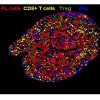
We demonstrate below a double immunostaining using a pair of rabbit antibodies to glucagon (amplified at a dilution of 1:5000 on path 12) and neurofilament M (conventionally stained at a dilution of 1:2000 on path 1) in a section of non-perfused rat pancreas. FITC-tyramide (green) is used in a simple fluorescent amplification (path 12) for the first antigen, glucagon, while Cy3-conjugated secondary antibody (red) is used in the conventional immunostaining (path 1) for the second antigen, neurofilament. We used the dilution of the first primary antibody, which is consistently not recognized by a conventional Cy3-labeled secondary antibody, but still clearly detectable by a simple fluorescent amplification method.
<center> <font></font> </center>
In addition to the controls described for the amplified immunofluorescent methods, we also used consecutive sections immunostained conventionally for either antigen separately. Furthermore, we selected antigen pairs for these double stainings which are known to be present in different cells. Thus, we could have easily recognized any cross-reaction between the first primary antibody and the second secondary antibody.
Use of TSA amplification in ISHH :
In effect, non-radioactive ISHH is the equivalent of an immunostaining after the hybridization in which the digoxigenin label of the probe is detected as an antigen in the tissue. Thus, after hybridization, the digoxigenin can be detected using the TSA amplification method that is described in detail in our
ISHH-TSA
protocol.
In our hands, riboprobes work very well this way, being almost as sensitive as radiolabelled riboprobes. Oligonucleotides have not worked as well so far using the TSA amplification method. The advantage of our method over biotin- or FITC-labelled probes is that the same digoxigenin probe can be used for a final product of alkaline phosphatase, DAB or fluorochrome, making double labelings easier.
Related Literature:
-
Adams JC (1992) Biotin amplification of biotin and horseradish peroxidase signals in histochemical stains. J Histochem Cytochem 40:1457-1463
-
Albertson, D.G., Fishpool, R.M. and Birchall, P.S. 1995. Fluorescence in situ hybridization for the detection of DNA and RNA.Methods Cell Biol 48:339-364.
-
Berghorn KA, Bonnett JH, Hoffman GE (1994) cFos immunoreactivity is enhanced with biotin amplification. J Histochem Cytochem 42:1635-1642
-
Bobrow MN, Harris TD, Shaughnessy KJ, Litt GJ (1989) Catalyzed reporter deposition, a novel method af signal amplification. Application to immunoassays. J Immunol Methods 125:279-285
-
Hsu SM, Raine L, Fanger H (1981) A comparative study of the peroxidase-antiperoxidase method and an avidin-biotin complex method for studying polypeptide hormones with radioimmunoassay antibodies. Am J Clin Pathol 75:734-738
Hunyady, B., Krempels, K., Harta, G. and Mezey, É (1996) Immunohistochemical signal amplification by catalyzed reporter deposition and its application in double immunostainings.J. Histochem. Cytochem. 44:1353-1362
-
Kerstens, H.M.J., Poddihe, P.J. and Hanselaar, A.G.J.M. (1995) A novel in situ hybridization signal amplification method based on the deposition of biotinylated tyramine.J. Histochem. Cytochem. 43:347-352.
-
Kiss A, Palkovits M, Skirboll LR (1988) Light microscopic triple-colored immunohistochemical staining on the same vibratome section using the avidin-biotin-peroxidase complex technique. Histochemistry 88:353-356
-
Negoescu A, Labat-Moleur F, Lorimier P, Lamarcq L, Guillermet C, Chambaz E, Brambilla E (1994) F(ab) secondary antibodies: a general method for double immunolabeling with primary antisera from the same species. Efficiency control by chemiluminescence. J Histochem Cytochem 42:433-437
-
Petrusz P, Ordronneau P, Finley JC (1980) Criteria of reliability for light microscopic immunocytochemical staining. Histochem J 12:333-348
- Wessel GM, McClay DR (1986) Two embryonic, tissue-specific molecules identified by a double-label immunofluorescence technique for monoclonal antibodies. J Histochem Cytochem 34:703-706
<center> <p> </p> </center>
上一篇:IMMUNOHISTOCHEMISTRY on PARAFFIN OR CRYOSECTIONS 下一篇:Immunohistochemistry