凋亡形态学图片
互联网
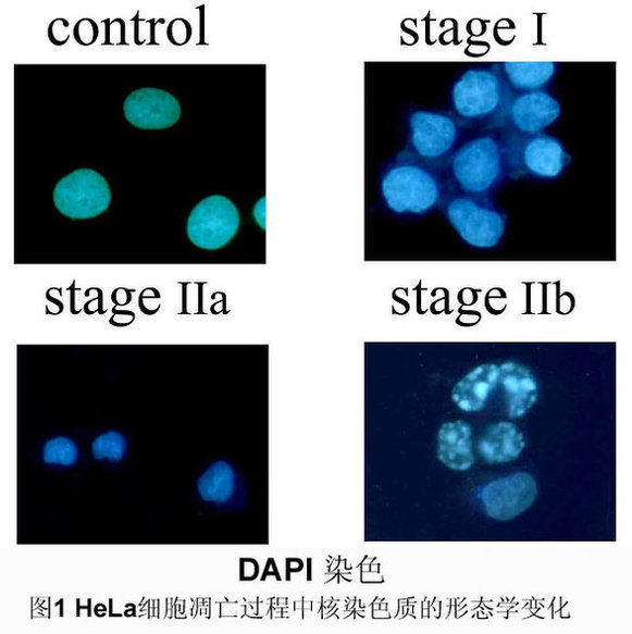
碧云天凋亡试剂盒hoechst33258染色,他们认为细胞发生凋亡时,染色质会固缩。 所以Hoechst染色时,细胞核会呈致密浓染,或呈碎块状致密浓染。
正常细胞核

刺激后有致密浓染的凋亡细胞

The image below shows human lymphoma cells treated with the chemotherapy agent camptothecin. The cells that are undergoing apoptosis appear yellow and show the characteristic membrane blebbing (bubble formation)seen in cells dying via apoptosis.

Promega公司的DeadEnd™比色法细胞凋亡检测系统能标记片断化的DNA,而这种片段化的DNA被视为是细胞凋亡的典型生化特征之一。
DeadEnd™比色法细胞凋亡检测系统是一种在组织切片与培养细胞中标记凋亡细胞核的理想方法,与此同时,还可以做形态学上的评估。本文中,将展示此种方法在细胞凋亡的原位细胞, 动物模型及多种病理学组织切片中的应用。
抗Fas单克隆抗体(50ng/ml;克隆CH-11,Oncor)诱导的Jurkat细胞的细胞凋亡。DeadEnd™比色法细胞凋亡检测系统标记Fas单克隆抗体处理过的细胞核(16小时),而未处理过的则未被染色(右下角插图)。

Apoptosis is of Greek origin meaning "falling off". The term is used in an analogy to the apparent suicide of leaves resulting in the very visible colour changes associated with the Autumn/Fall and that eventually leads to the leaves falling from the trees. Similiarly, cells go through a preordained sequence of events resulting in death and removal from the body.
Apoptosis is a regimented sequence of events, with each act being executed in a timed fashion. Its orderliness is often referred to as the "dance of death". Microscopic examination of the cell reveals that there are events occurring both externally and internally that can be listed as follows:
Externally:
Shrinkage or loss of volume of the cell
Blebbing on the surface of the cell
Externalisation of a phospholipids termed phosphatidylserine
Internally:
Condensation of cytoplasm
Breakdown in mitochondria integrity with the subsequent release of apoptosis inducing factors such as cytochrome c
Activation of caspases
Degradation of chromosomal DNA into 180-bp internuclosomal fragments
Degradation of a large number of proteins (>100) thought to be important in the cell survival and metabolism
Two human tumour cells, one normal (left) and the second (right) undergoing apoptosis following exposure to cytotoxic drugs
DeadEnd™比色法细胞凋亡检测系统实验流程:

What happens during apoptosis?
To kill itself, a cell first makes a deadly chemical cocktail. It then separates itself from its neighbours, and unleashes the poisons. These include a substance that chews up the DNA in the cell nucleus, and a 'glue' that binds the inside of the cell together. Within a few hours, the cell shrinks, breaks up and is engulfed by other cells.
A cancer cell (mauve) undergoing apoptosis.
Dr Andrejs Liepins/Science Photo Library

Apoptotic human keratinocytes that were fixed with paraformaldehyde and stained with fluorescein concanavalin A (C827). The cells were subsequently permeabilized with acetone, stained with propidium iodide (P1304, P3566, P21493) and visualized by confocal laser-scanning microscopy. This photomicrograph clearly shows that the green-fluorescent lectin staining outlines the apoptotic surface blebs, whereas the red-fluorescent propidium iodide stains the interior of the blebs. Image contributed by Livia Casciola-Rosen and Antony Rose, Johns Hopkins University. Reproduced from by copyright permission of the Rockefeller University Press.
凋亡的人类角化细胞被多聚甲醛固定,被荧光素标记的刀豆蛋白A染色。然后经丙酮渗透处理,被pi染色后能被共聚焦显微镜观察。本片显示的绿色荧光(外源性凝集素)勾勒出凋亡细胞表面的小泡,红色荧光(pi)被包被在其中。

Apoptosis induced in Jurkat cells with 10 µM camptothecin. The cells were then treated with the reagents in the Vybrant Apoptosis Assay Kit #4 (V13243). Apoptotic cell nuclei were labeled with YO-PRO-1 dye (green) (Y3603). Necrotic cells were detected with propidium iodide (red)
10 µM喜树碱诱导的Jurkat cells(慢淋。G1检测点缺陷细胞) 凋亡。细胞然后经 Vybrant Apoptosis Assay Kit #4 (V13243).处理。凋亡细胞核被YO-PRO-1绿染,坏死细胞被pi红染。

Jurkat human T-cell leukemia cells treated with 1 µM camptothecin. The externalized phosphatidylserine, a characteristic of early-stage apoptotic cells, was detected with Alexa Fluor 488 annexin V (A13201). The late-stage apoptotic and necrotic cells were stained with propidium iodide (P1304, P3566, P21493). The image was acquired using bandpass filters appropriate for fluorescein.
Jurkat 人类白血病T细胞经1微升喜树碱处理,ps外翻,早期凋亡特异性的特征被annexin-5检测到。晚期凋亡和坏死细胞被pi染色。本图是由相应的荧光滤光片获取的。

HL-60 cells treated with camptothecin for three hours. The DNA strand nicks characteristic of apoptosis were detected with the TUNEL (terminal deoxynucleotidyl transferase–mediated dUTP nick end-labeling) assay using the fluorescently labeled nucleotide, ChromaTide BODIPY FL-14-dUTP (C7614). Image contributed by Zbigniew Darzynkiewicz, Cancer Research Institute, New York Medical College.
HL-60 cells(急粒,p53缺陷)被喜树碱诱导3小时。DNA段端被荧光标记的脱氧核苷酸末端转移酶标记。

Detection of apoptosis in SK-N-MC neuroblastoma cells. Following a six-hour exposure to hydrogen peroxide, cells were labeled with Hoechst 33342 (H1399, H3570, H21492), tetramethylrhodamine ethyl ester (TMRE, T669) and rhodamine 110, bis-L-aspartic acid amide (R22122) for 15 minutes. Apoptotic cells show green cytosolic fluorescence resulting from cleavage of the rhodamine 110, bis-L-aspartic acid amide substrate by active caspase-3. The staining pattern of the Hoechst 33342 dye reveals that the majority of the rhodamine 110–positive cells also contain condensed or fragmented nuclei characteristic of apoptosis. Furthermore, the rhodamine 110–positive cells are also characterized by an absence of polarized mitochondria, as indicated by their failure to load the positively charged mitochondrial indicator TMRE. The image was contributed by A.K. Stout and J.T. Greenamyre, Emory University.

线虫发育中细胞的凋亡:
用共聚焦显微镜观察线虫胚胎,在正常胚胎CED-4位于线粒体。而凋亡细胞CED-4(红色)和核纤层蛋白(绿色)在核被膜(黄色)均可见。该实验有助于揭示,在细胞凋亡中CED-4从线粒体易位到核被膜