Static and Dynamic Imaging of Erythrocyte Invasion and Early Intra-erythrocytic Development in Plasmodium falciparum
互联网
互联网
相关产品推荐

Mitochondrial dynamic protein MID51, Smcr7l ELISA KIT/ Mouse Mitochondrial dynamic protein MID51, Smcr7l ELISA试剂盒
¥2980

US12/US12蛋白Recombinant Human herpesvirus 1 ICP47 protein (US12)重组蛋白Immediate-early protein IE12Immediate-early-5Infected cell protein 47US12 proteinVmw12蛋白
¥2616

iagB/iagB蛋白Recombinant S_a_l_monella t_y_p_himurium Invasion protein iagB (iagB)重组蛋白iagB; STM2877Invasion protein IagB蛋白
¥2328

Intra Acrosomal Protein Rabbit pAb(bs-7866R)-50ul/100ul/200ul
¥1180

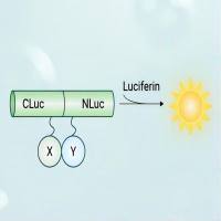
荧火素酶互补实验(Luciferase Complementation Assay, LCA)| 荧光素酶互补成像技术(Luciferase Complementation Imaging, LCI)
¥5999
相关问答

