Quantitative Imaging of Chemical Composition in Single Cells by Secondary Ion Mass Spectrometry: Cisplatin Affects Calcium Stores in Renal Epithelial
互联网
572
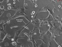
A detailed protocol for quantitative single cell mass spectrometry imaging (MSI) analysis is described in this chapter with examples of the treatment of cells with anticancer drug, cisplatin. Cisplatin, cis -diamminedichloridoplatinum ii (CDDP), is widely used for the treatment of many malignancies, including testicular, ovarian, bladder, cervical, head and neck, and small cell and non-small cell lung cancers. The possibility of renal injury by cisplatin treatment is a major dose-limiting factor in this cancer therapy. At present, the mechanisms of cisplatin-induced renal cytotoxicity are poorly understood. In this work, secondary ion mass spectrometry (SIMS) was used for investigating cisplatin-induced alterations in intracellular chemical composition in a well-established model (LLC-PK1 cell line) for studying renal injury. The cells were cryogenically prepared by the sandwich freeze-fracture method for subcellular imaging analysis of chemical composition (total concentrations of K+ , Na+ , and Ca2+ ) in individual cells. The single cell analysis of these diffusible ions necessitates the use of reliable cryogenic sample preparations for SIMS. The sandwich freeze-fracture method offers a simple approach for cryogenically preserving diffusible ions and molecules inside the cells for SIMS analysis. A CAMECA IMS-3f SIMS ion microscope instrument capable of producing chemical images of single cells with 500-nm spatial resolution was used in the study. In cisplatin-treated cells, SIMS imaging showed the presence of detectable amount of platinum at mass 195, as 195 Pt+ secondary ions in individual cells. SIMS observations also revealed that individual cells differed in their response to cisplatin. While the chemical composition of some cells was unaffected by cisplatin, others showed a reduction in cytoplasmic calcium stores that was not associated with changes in their intracellular K or Na concentrations. Another population of cells displayed an increase in cytoplasmic calcium concentration that was associated with higher levels of intracellular Na and a reduction in K concentration of the same cells. Since the loss of intracellular K and the gain of Na and Ca are typical symptoms of cell injury, it is plausible that the initial response of the cell to cisplatin treatment is the reduction in cytoplasmic calcium pool in stores. If, somehow, the calcium stores are compromised with cisplatin, then maintenance of free Ca2+ homeostasis would become uncontrollable in the cell. These observations open new avenues of research for understanding of the mode of action of cisplatin in cell injury. This study also demonstrates the need and vast potential of single cell imaging mass spectrometry techniques in cell biology and medicine.