Slide Preparation for Laser Capture Microdissection (LCM)
互联网
These methods were successful in our lab using prostate tissue and for our specific objectives. Investigators must be aware that they will need to tailor the following protocol for their own research objectives and tissue under study.
LCM and subsequent molecular analysis can be carried out on slides stained using standard hematoxylin and eosin methods. However, if cell types that are (or are not) expressing a specific protein are required for a study then more advanced slide preparation methods such as Immuno-LCM may be utilized.
1. Materials
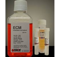
-
- 70%, 95%, 100% ethanol
- Xylenes, mixed, ACS reagent (Sigma)
- Deionized water
- Hematoxylin solution, Mayer's (Sigma)
- Eosin Y solution (Sigma)
- Complete, mini protease inhibitor cocktail tablets (Roche Corp.)
Important: For all protein analysis, dissolve 1 protease inhibitor cocktail tablet per 10 ml of each reagent except the xylenes.
2. Storage of Sections
- Recut paraffin sections are stored at or below room temperature. Do not deparaffinize until immediately prior to microdissection.
- Low-melt polyester sections are stored at 4°C.
- Frozen sections are stored at -80°C or below.
3. Methods
TIP: Use the minimal amount of staining to visualize the tissue for microdissection. This will significantly improve macromolecule recovery. For example, hematoxylin and eosin can be used at 10% of their standard concentrations. Since the slides are microdissected without a coverslip, the tissue is not index-matched and substantial light scattering occurs, typically producing "dark" images. Thus, both image quality and molecular recovery can be improved by decreasing stain concentrations.
A: Paraffin-embedded Sections or Frozen Sections
- If a paraffin-embedded section is to be stained, start from step 1.
- If the section was frozen-embedded, melt it gently (e.g., on the back of the hand) for approximately 30 sec after removal from the freezer. This will create a "rougher" tissue surface and allow for better adhesion to the LCM cap. Start at Step 4.
Place the sections in the following solutions:
-
- Fresh xylenes (to depariffinize the sections) - 5 min
- Fresh xylenes - 5 min
- 100% ethanol - 15 sec
- 95% ethanol - 15 sec
- 70% ethanol - 15 sec
- Deionized water - 15 sec
- Mayer's Hematoxylin - 30 sec
- Deionized water - rinse (x 2) - 15 sec
- 70% ethanol - 15 sec
- Eosin Y - 5 sec
- 95% ethanol - 15 sec
- 95% ethanol - 15 sec
- 100% ethanol - 15 sec
- 100% ethanol - 15 sec
- Xylenes (to ensure dehydration of the section) - 60 sec
- Air-dry for approximately 2 minutes or gently use air gun to completely remove xylenes.
- The tissue is now ready for LCM.
B: Low-melt Polyester-embedded Sections
TIP: Proceed gently when staining sections embedded in polyester wax. Even though the sections are placed on charged slides, the tissue has a tendency to detach from the slide and should be monitored carefully throughout the staining procedure.
Place the sections in the following solutions:
-
- 100% ethanol (to remove polyester wax) - 5 min
- 100% ethanol - 5 min
- 95% ethanol - 15 sec
- 70% ethanol - 15 sec
- Deionized water - 15 sec
- Mayer's hematoxylin - 30 sec
- Deionized water - 15 sec
- 70% ethanol - 30 sec
- Eosin Y - 5 sec
- 95% ethanol - 15 sec
- 95% ethanol - 15 sec
- 100% ethanol - 15 sec
- 100% ethanol - 15 sec
- 50:50, xylenes:100% ethanol - 10 sec
- The tissue is now ready for LCM .
TIP: The xylenes-ethanol step at the end of the procedure is critical for subsequent LCM. The length of time may need to be adjusted depending on the tissue type and goals of the study. For example, if the tissue is left in this solution longer than 10-15 sec, the tissue may detach from the slide during dissection. Conversely, if the xylenes-ethanol step is too short, the tissue may be strongly bound to the slide and not dissect well.
TIP: The tissue section should be completely dry before LCM. Use of an Accuduster or similar device may facilitate drying for efficient microdissection.