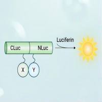
A number of altered pathways in cancer cells depend on growth factor receptors. The amplification/alteration of the epidermal growth factor receptor (EGFR) has been shown to play a significant role in enhancing tumor burden in a number of tumors, including malignant glioblastomas (GBM). To dissect the role of EGFR expression in tumor progression in mouse models of cancer and ultimately evaluate targeted therapies, it is necessary to visualize the dynamics of EGFR in real time in vivo. Non-invasive imaging based on quantitative and qualitative changes in light emission by fluorescent and bioluminescent markers offers a huge potential to facilitate drug development. Multiple approaches could be used to follow a molecular target or pathway with the fusion of a bioluminescent–fluorescent marker. This unit describes a protocol for simultaneously imaging EGFR activity and progression of GBM in a mouse model. Human glioma cells transduced with lentiviral vectors bearing different combinations of fluorescent and bioluminescent proteins either fused to EGFR or expressed alone can be grown as monolayers and maintained over several passages. The unit begins with a method for transducing glioma cells with lentiviral vectors for stable expression of these fluorescent and bioluminescent markers in vitro, followed by transplantation of engineered glioma cells in mice, and, finally, sequential bioluminescent imaging of EGFR expression and GBM progression in mice. The protocol details characterization of engineered glioma cells in culture, surgical preparation, craniotomy, cell implantation, animal recovery, and imaging procedures to study kinetics of EGFR expression and GBM progression.


