Imaging Sea Urchin Fertilization
互联网
625
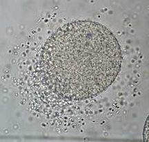
Imaging animal fertilization presents a number of unique challenges that arise from the unusual nature of the cells involved. Eggs are among the largest cells produced by animals, and sperm are frequently the smallest. Observation of the initial steps of fertilization, including sperm-egg binding and fusion, can be challenging owing to the size discrepancy between these two cell types and, in many species, the rapid time course over which they occur. Subsequent events in fertilization, including the remarkable activities of the microtubular cytoskeleton during the first cell cycle, are usually obscured by the mass of the egg cytoplasm, and only the most obvious features, such as pronuclear formation, are apparent by routine microscopic methods. The application of confocal microscopy methods to the demands of imaging studies in fertilization has proven to be an extremely valuable approach, as the morphological features of most types of animal eggs are extremely well suited to the specific advantages this technology offers. Confocal microscopy is a powerful and flexible tool, and has been skillfully used and developed by a number of researchers not only to study the structural features of fertilization at high resolution, but also to examine dynamic events in living gametes, including imaging signaling events that occur during and after gamete fusion.