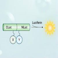
Luciferase Protein Complementation Assays for Bioluminescence Imaging of Cells and Mice
互联网
互联网
相关产品推荐

WIP (Lysis Buffer for WB/IP Assays, Nondenaturing)(C-0013)-30ml/60ml/100ml
¥70

Coronavirus Nucleocapsid重组蛋白|Recombinant SARS-CoV-2 Nucleocapsid-AVI&His recombinant Protein,Biotinylated
¥4520

///蛋白/P-Mice-Ins-betundefined NaTx5.4蛋白/Recombinant Tityus bahiensis Toxin Tb2-II重组蛋白
¥69

MKN45人低分化胃癌细胞|MKN45细胞(Human Poorly Differentiated Gastric Cancer Cells)
¥1500

荧火素酶互补实验(Luciferase Complementation Assay, LCA)| 荧光素酶互补成像技术(Luciferase Complementation Imaging, LCI)
¥5999
相关问答

