Separation of sea urchin blastomeres
互联网
Objective: to separate the Blastomere at different points in development and observe the growth of the resulting embryos.
Procedure: We extractrf the gametes from the sea urchins, fertilized the sea urchin eggs, and removed the fertilization envelope. The cells were then be allowed to cleave, at which point they were be introduced into a hypertonic seawater solution. This caused the cells to shrink and break apart. Once the majority of the cells had separated, the water was replaced with normal seawater. Their growth was be compared to that of the control group that was not divided.
- The eggs were washed several times with ASW by allowing them to settle and pouring off the water. This will removed some of the jelly coat.
- Enough eggs to cover the bottom of a 50 ml beaker were transfered along with 10 ml of 10mM p-aminobenzoic acid in ASW.
- 50 ml of sperm were diluted in 5 ml of ASW. 2 drops were used to fertilize eggs.
- The presence of the fertilization envelope was checked after 5 and 10 minutes in the control culture.
- The eggs were poured through a nylon filter mounted to the end of a 50 ml beaker. This was repeated until most of the fertilization envelopes have been removed.
- The cells were allowed to settle for a few minutes.
Blastomere Separation
- The cells were then separated into 3 100 ml beakers full of ASW. Care was taken so as not to add too much of the seawater-PABA solution.
- We then waited for the control culture to cleave for the first time.
- Then most of the water was poured from the first experimental culture.
- The beaker was gently filled with hypertonic seawater. The culture was then checked after 5 minutes to see whether osmotic shrinkage caused the cells to separate. The solution was replaced with additional hypertonic seawater if they had not separated.
- We waited 10 minutes after the cells show osmotic shrinkage.
- The hypertonic solution was poured off and replaced it with normal seawater. This was repeated.
- Steps 3-6 were repeated for the second experimental culture once the control group cleaved a third time.
- The embryos were allowed to develop and observed at different stages in their growth.
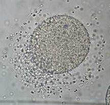
Results
While the control embryos grew to form normal pluteus the experimental embryos did not cleave once seperated from each other. The control organisms were stained with 5C7, EctoV, and Ig8 to look at their formation even though there were no experimental clones to compare to them. Another procedure involving a calcium poor seawater solution can be used to seperate the embryos and may produce more desirable results.
Click on thumbnails to see full size pictures.
Use the back button of your browser to return to this page