溶组织内阿米巴 Entamoeba histolytica
互联网
| Introduction |
|
<center>
<table>
<tbody>
<tr>
<td>Kingdom</td>
<td>Subkingdom</td>
<td>Phylum</td>
<td>Class</td>
<td>Order</td>
<td>Family</td>
<td>Genus+Species</td>
</tr>
<tr>
<td>Protista</td>
<td>Protozoa</td>
<td>sarcomastigophora</td>
<td>Lobosea</td>
<td>Amoebida</td>
<td>Entamoebidae</td>
<td>Entamoeba Histolytica</td>
</tr>
</tbody>
</table>
</center>
|
|
Morphology
Pay your attention to stages that have diagnostic value
|
| 1. cyst (non-motile) |
| (1) 10-20 mocrometers in size |
| (2) spherical in shape |
| (3) 1-2 nuclei (immature cyst); 4 nuclei (mature cyst-infective stage). |
|
(4) inclusions:(become smaller and smaller as the cyst ages)
glycogen vacuole appears as a clear space; food reservoir chromatoid body dark blue rods or dots; its function is not known |
| 2. trophozoite (active form) |
| (1) Size : 10-40 micrometers in diameter, some are above 60 micrometers. |
|
(2) Pseudopodium
(ectopalsmic protrusion):
A broad or finger-like in form B thrust out quickly C firstly, formed with ectoplasm, secondly, endoplasm flows slowly into it. D motility is progressive and directional. |
| (3) Endoplasm : red blood cells may be found in it. |
|
(4) Nucleus
(vesicular type)
It is not visible in an unstained specimen; but its clear structure can be seen when stained with hematoxylin. A: membrane: distinct line B: chromatin granules: fine ane uniformally arranged in the inner surface of the nulear membrane. C: karyosome: small and centrally located. |
|
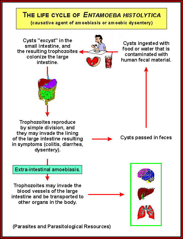
Life Cycle |
|
|
| 1 infective stage: mature cyst |
| 2 access: mouth |
| 3 ecological niches: large intestine; liver, lung and other organs. |
| 4 pathogenic stage: trophozoite |
| 5 diagnostic stage: cyst; trophozoites |
|
Pathogenesis |
|
一、Clinical classification
Asymptomatic infection (carrier)>90% cases sympomatic cases<10% A. Intestinal amoebiasis a. dysentery b. non-dysenteric colitis c.appendicitis d.amoeboma B. Extra-intestinal amoebiasis a. Hepatic (1) acute non-suppurative (2) liver abscess b. Pulmonary c. Brain d. Skin e. Other extra-intestinal amoebiasis 二、Pathogenic factors 1.toxicity of parasites pathogenic-nonpathogenic complex.
Entamoeba histolytica
2.symbiotic bacteria 3.defence barrier immunity 三、pathology pinpoint lesion on mucous membrane flask-shaped crateriform ulcers Diagosis
<center>
<table>
<tbody>
<tr>
<td></td>
<td>
<p>trophozoite</p>
</td>
<td>
<p>cyst</p>
</td>
</tr>
<tr>
<td>specimen</td>
<td>
<p>feces</p>
</td>
<td>
<p>feces</p>
</td>
</tr>
<tr>
<td>
<p>method</p>
</td>
<td>direct smear with normal saline</td>
<td>direct smear with iodine stain</td>
</tr>
<tr>
<td>
<p>diseases</p>
</td>
<td>amoebic dysentery</td>
<td>chronic intestinal amoebiasis or carriers</td>
</tr>
<tr>
<td>
<p>remarks</p>
</td>
<td>
1.container must clean
<br />
2.examined soon after they have been passed.
<br />
3.select bloody and mucous portion.
</td>
<td></td>
</tr>
</tbody>
</table>
</center>
Epidemiology Distribution: tropical and subtropical areas. Media: flies; black beetles etc. Treatment and prevention treatment: diodoquin-carriers metronidazole-dysentery prevention human feces should not be used as fetilizer food and drinks must be protected from flies personal hygiene: wash hands after defecation and before meals. |