

- ¥7500
- GeneTex

- 美国
- GTX02575-pro
- 2025年07月12日

企业认证
相关产品推荐更多 >
万千商家帮你免费找货
0 人在求购买到急需产品
- 详细信息
- 技术资料
- 保存条件:
Store as concentrated solution. Centrifuge briefly prior to opening vial. Aliquot and store at -20ºC or below. Avoid multiple freeze-thaw cycles.
- 保质期:
12 months from the shipping date of the product.
- 英文名:
SARS-CoV-2 (COVID-19) Spike (D614G Mutant)(ECD) protein, His tag (active)
- 库存:
Available
- 供应商:
GeneTex
- 规格:
100 μg
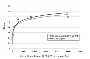
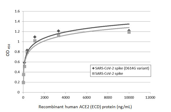
Functional ELISA analysis of immobilized recombinant SARS-CoV-2 (COVID-19) Spike (D614G variant) protein, His tag (active) (GTX02575-pro), and SARS-CoV-2 spike (trimer) protein (coated at 2 μg/mL) binding to soluble recombinant Human ACE2 (ECD) protein , mouse IgG Fc tag (GTX135683-pro) (13-10000 ng/mL). Bound protein was detected by Goat Anti-Mouse IgG antibody (HRP) (13111-01) (1:10000).
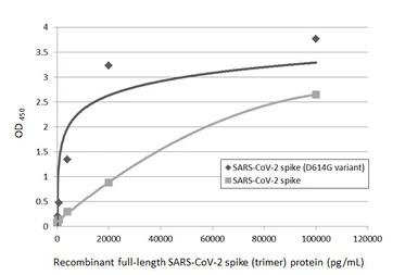
Sandwich ELISA detection of recombinant SARS-CoV-2 (COVID-19) Spike (D614G variant) protein, His tag (active) (GTX02575-pro), and SARS-CoV-2 spike (trimer) protein using SARS-CoV-2 (COVID-19) Spike RBD antibody [HL1014] (GTX635807) as capture antibody at concentration of 5 μg/mL and SARS-CoV-2 (COVID-19) Spike RBD antibody [HL1003] (HRP) (GTX635792-01) as detection antibody at concentration of 1 μg/mL.
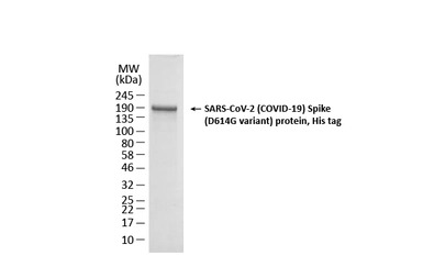
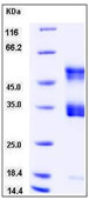
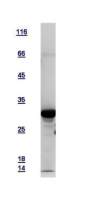
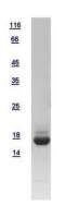
SDS-PAGE of GTX02575-pro SARS-CoV-2 (COVID-19) Spike (D614G variant) protein, His tag.
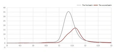
Gel filtration analysis of 2 different batches of spike protein.
The first batch : spike protein in PBS buffer.
The second batch : spike protein in PBS, 20% glycerol.
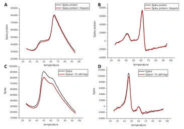
Differential scanning fluorimetry (DFS) analysis the stability and the heparin binding ability. DSF of proteins were performed in the absence or presence of heparin (10 μM). The scanning fluorimetry data of the two batches shows the protein is both stable and functional, with heparin binding activity.
A and C : Melting curve of spike protein.
B and D : First derivative of the melting curves of spike to show its melting temperature as peak.
A and B : the first batch of spike protein (PBS)
C and D : the second batch of spike protein (PBS, 20% glycerol).
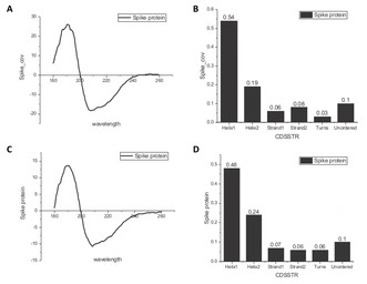
CD spectra of different cation forms of spike protein. The scanning fluorimetry data of the two batches shows the protein is both stable and functional, with heparin binding activity.
A and C : CD spectra were recorded on J-1100 spectrometer between 180 and 260 nm of spike protein (0.5 mg/ml).
B and D : Secondary structure were analysed by program CDSSTR of A and C, respectively.
A and B : the first batch of spike protein (PBS buffer).
C and D : the second batch of spike protein (PBS, 20% glycerol).
风险提示:丁香通仅作为第三方平台,为商家信息发布提供平台空间。用户咨询产品时请注意保护个人信息及财产安全,合理判断,谨慎选购商品,商家和用户对交易行为负责。对于医疗器械类产品,请先查证核实企业经营资质和医疗器械产品注册证情况。
 技术资料
技术资料暂无技术资料 索取技术资料

